Deck 22: Stem Cells and Tissue Renewal
Full screen (f)
Question
Question
Question
In the 1950s, scientists fed  o rats to label cells that were synthesizing DNA, and then followed the fates of labeled cells for periods of up to a year. They found three patterns of cell labeling in different tissues. Cells in some tissues such as neurons in the central ner- vous system and the retina did not get labeled. Muscle, kidney, and liver, by contrast, each showed a small number of labeled cells that retained their label, apparently with- out further division or loss. Finally, cells such as those in the squamous epithelia of the tongue and esophagus were labeled in fairly large numbers, with radioactive pairs of nuclei visible in 12 hours; however, the labeled cells disap- peared over time. Which of these three patterns of labeling would you expect to see if the labeled cells were generated by stem cells? Explain your answer.
o rats to label cells that were synthesizing DNA, and then followed the fates of labeled cells for periods of up to a year. They found three patterns of cell labeling in different tissues. Cells in some tissues such as neurons in the central ner- vous system and the retina did not get labeled. Muscle, kidney, and liver, by contrast, each showed a small number of labeled cells that retained their label, apparently with- out further division or loss. Finally, cells such as those in the squamous epithelia of the tongue and esophagus were labeled in fairly large numbers, with radioactive pairs of nuclei visible in 12 hours; however, the labeled cells disap- peared over time. Which of these three patterns of labeling would you expect to see if the labeled cells were generated by stem cells? Explain your answer. Question
At any given time, intestinal crypts of mice com- prise about 15 stem cells and 10 Paneth cells. After cell division, which occurs about once a day, the daughter cells remain stem cells only if they maintain contact with a Paneth cell. This constant competition for Paneth-cell con- tact raises the possibility that crypts might become mono- clonal over time; that is, the crypt cells at one point in time might derive from only 1 of the 15 stem cells that existed at some earlier time. To test this possibility, you use the so-called confetti marker that upon activation expresses any one of three fluorescent proteins in the stem cells of the crypt. You then examine crypts at various times to determine whether they contain cells with multiple colors or only one color (Figure Q22-1). Do the crypts become monoclonal over time or not? How can you tell? 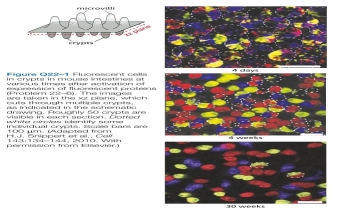
Question
The origin of new  cells of the pancreas-from stem cells or from preexisting
  cells-was not resolved until a decade ago, when the technique of lineage tracing was used to decide the issue. Using transgenic mice that expressed a tamoxifen-activated form of Cre recombinase under the control of the insulin promoter, which is active only in
  cells, investigators could remove an inhibitory segment of DNA and thereby allow expression of human placental alkaline phosphatase (HPAP), which can be detected by histochemical staining. After a pulse of tamox- ifen that converted about 30% of
  cells in young mice to cells that express HPAP, the investigators followed the per- centage of labeled
  cells for a year, during which time the total number of
  cells in the pancreas increased by 6.5- fold. How do you suppose the percentage of
  cells would change over time if new
  cells were derived from stem cells? What if new
  cells were derived from preexisting
  cells? WFigure Q22.03/Q22.01hich hypothesis do the results in Figure Q22-2 support?
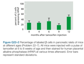
cells of the pancreas-from stem cells or from preexisting cells-was not resolved until a decade ago, when the technique of lineage tracing was used to decide the issue. Using transgenic mice that expressed a tamoxifen-activated form of Cre recombinase under the control of the insulin promoter, which is active only in cells, investigators could remove an inhibitory segment of DNA and thereby allow expression of human placental alkaline phosphatase (HPAP), which can be detected by histochemical staining. After a pulse of tamox- ifen that converted about 30% of cells in young mice to cells that express HPAP, the investigators followed the per- centage of labeled cells for a year, during which time the total number of cells in the pancreas increased by 6.5- fold. How do you suppose the percentage of cells would change over time if new cells were derived from stem cells? What if new cells were derived from preexisting cells? WFigure Q22.03/Q22.01hich hypothesis do the results in Figure Q22-2 support? Question
Question
Question
Question
Which statements are true? explain why or why not.
0 Generation of induced pluripotent stem (iPS) cells was first accomplished using retroviral vectors to carry the OSKM set of tran- scription regulators into cells. The efficiency of fibroblast reprogramming was typically low (0.01%), in part because large numbers of retroviruses must integrate to bring about reprogramming and each integration event carries with it the risk of inappropriately disrupting or activating a critical gene. In what other ways, or other forms, do you suppose you might deliver the OSKM transcription regula- tors so as to avoid these problems?
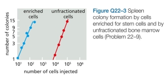
0 Generation of induced pluripotent stem (iPS) cells was first accomplished using retroviral vectors to carry the OSKM
set of tran- scription regulators into cells. The efficiency of fibroblast reprogramming was typically low (0.01%), in part because large numbers of retroviruses must integrate to bring about reprogramming and each integration event carries with it the risk of inappropriately disrupting or activating a critical gene. In what other ways, or other forms, do you suppose you might deliver the OSKM transcription regula- tors so as to avoid these problems? Question

Unlock Deck
Sign up to unlock the cards in this deck!
Unlock Deck
Unlock Deck
1/10
Play
Full screen (f)
Deck 22: Stem Cells and Tissue Renewal
1
Which statements are true? explain why or why not.
Every tissue that can be renewed is renewed from a tissue-specific population of stem cells.
Every tissue that can be renewed is renewed from a tissue-specific population of stem cells.
Osteoporosis is a bone disorder characterized by the thinning of the bone. The bone becomes highly porous and fragile. The bones appear like a honey comb under the microscope. The bones can become extremely painful and may make the locomotion of the patients difficult. Osteoblasts are the cells that secrete the substance for the bone formation. These are uninucleate. These cells synthesize collagen. Collagen is a network or mesh which connects the bones. Osteoclasts are also the bone cells. They are uninucleate. These Osteoclasts break down the bone tissues. This helps in the maintenance, repair and repair of the bone. They secrete collagenase and digest the protein and the mineral of the bone. The osteoclast and the osteoblast together function in an equilibrium to maintain healthy bones. The mismatch in the coordination of the osteoclast and the osteoblasts can give rise to osteoporosis. The bone forming tendency of the osteoblasts decreases, while the osteoclast keeps on breaking the bone. As a result, the misbalance causes the thinning of the bone. So, the given statement is  .
.
. 2
Disturbance of the balance in the activities of osteoblasts and osteoclasts in favor of osteoclasts can give rise to the condition known as osteoporosis, the brit- tle-bone syndrome of the elderly. Discuss the following problems.
Radioactive thymidine or thymidine analog gets incorporated in the DNA (deoxyribonucleic acid) of those cells which are undergoing S phase or cell division. These cells as they divide further, the thymidine label get diluted halving after every cell division. This can be quantifies in tissues. All cells present in an organism cannot repair and renew. Many cells like neurons or sensory epithelia of ear are terminally differentiated and are permanently lost on damage. They cannot be replenished. Some cells self renew but not depending on stem cells. Cells like ?-cells of pancreas or hepatocytes of liver continuously divide themselves without the need for stem cells. Their rate of division is very slow and division includes making a copy of them. Lining of gut continuously and rapidly renew itself but with the help of stem cells. When scientists fed radioactive thymidine to rats, three labeling pattern were seen. Their conclusion has been discussed as follow- 1. Neurons and retina did not get labeled - Radioactive thymidine can label only those cells which divide and are in S phase. No labeling took place in neurons or retina simply concludes that these cells were not dividing.
2. Muscles, kidney and liver showed slight labeling that retained the label with no further division - Inclusion of thymidine in these cells shows that cells went on a single cellular division when the thymidine got incorporated into its cells. But labeling was retained and radioactivity was not halved shows that no further division took place. This concludes that this type of division took place without the support of stem cells. Cells underwent the division on their own. 3. Squamous epithelial of tongue and esophagus labeled in large number with radioactive pairs of nuclei visible in 12 hours and labeled cells disappeared over time - Widespread labeling shows high cellular division with a cell cycle time of 12 hours as after which cells underwent another round of cellular division. The originally labeled cell got disappeared over time. This observation comes in close to the type of renewal of cells taking place with the help of stem cells. Stem cells divide rapidly and the two daughter cells can choose their own fate. Fate to remain a stem cell or to differentiate further into terminally differentiated cells. Loss of labeling over time hints towards that cells after labeling and getting differentiated underwent apoptosis to give way to new other cells. Hence, the last pattern showed by squamous epithelial and esophagus shows the pattern of labeled stem cell.
2. Muscles, kidney and liver showed slight labeling that retained the label with no further division - Inclusion of thymidine in these cells shows that cells went on a single cellular division when the thymidine got incorporated into its cells. But labeling was retained and radioactivity was not halved shows that no further division took place. This concludes that this type of division took place without the support of stem cells. Cells underwent the division on their own. 3. Squamous epithelial of tongue and esophagus labeled in large number with radioactive pairs of nuclei visible in 12 hours and labeled cells disappeared over time - Widespread labeling shows high cellular division with a cell cycle time of 12 hours as after which cells underwent another round of cellular division. The originally labeled cell got disappeared over time. This observation comes in close to the type of renewal of cells taking place with the help of stem cells. Stem cells divide rapidly and the two daughter cells can choose their own fate. Fate to remain a stem cell or to differentiate further into terminally differentiated cells. Loss of labeling over time hints towards that cells after labeling and getting differentiated underwent apoptosis to give way to new other cells. Hence, the last pattern showed by squamous epithelial and esophagus shows the pattern of labeled stem cell.
3
In the 1950s, scientists fed o rats to label cells that were synthesizing DNA, and then followed the fates of labeled cells for periods of up to a year. They found three patterns of cell labeling in different tissues. Cells in some tissues such as neurons in the central ner- vous system and the retina did not get labeled. Muscle, kidney, and liver, by contrast, each showed a small number of labeled cells that retained their label, apparently with- out further division or loss. Finally, cells such as those in the squamous epithelia of the tongue and esophagus were labeled in fairly large numbers, with radioactive pairs of nuclei visible in 12 hours; however, the labeled cells disap- peared over time. Which of these three patterns of labeling would you expect to see if the labeled cells were generated by stem cells? Explain your answer.
o rats to label cells that were synthesizing DNA, and then followed the fates of labeled cells for periods of up to a year. They found three patterns of cell labeling in different tissues. Cells in some tissues such as neurons in the central ner- vous system and the retina did not get labeled. Muscle, kidney, and liver, by contrast, each showed a small number of labeled cells that retained their label, apparently with- out further division or loss. Finally, cells such as those in the squamous epithelia of the tongue and esophagus were labeled in fairly large numbers, with radioactive pairs of nuclei visible in 12 hours; however, the labeled cells disap- peared over time. Which of these three patterns of labeling would you expect to see if the labeled cells were generated by stem cells? Explain your answer.Intestinal crypts consist of both stem cells and paneth cells. All the cells of the crypt can rise from a single stem cell. Paneth cells via Wnt (wingless-type mouse mammary tumor virus) signaling maintain the undifferentiated state of the cells in close contact with them. It was found that there are 15 stem cells and 10 paneth cells in a crypt. It was suspected that it may happen that all the cells that might be present in the crypt at a particular point may have been produced from the same single stem cell, which went into multiple division producing differentiated cells. To test this hypothesis, different stem cells present in the crypt were marked with different fluorescent protein linked to the same type of protein being expressed in all stem cells. This fluorescence was followed for a few weeks to know if all the cells could have been derived from the same stem cell. The results were observed as follows: 1. On the 4 th day after labeling, one can see 3 different fluorescent proteins in a single crypt-blue, yellow-green and red.
2. On the 4 th week, one can see that most of the crypts have a single color- yellow or red or blue. 3. On the 30 th week, many of the crypts lose their fluorescence, but the rest crypts show a single color- blue or red or yellow. Yes, at some point of time, only a single stem cell present in the crypt is responsible for the differentiation of all the cells of the crypt. Hence, the crypt becomes monoclonal.
2. On the 4 th week, one can see that most of the crypts have a single color- yellow or red or blue. 3. On the 30 th week, many of the crypts lose their fluorescence, but the rest crypts show a single color- blue or red or yellow. Yes, at some point of time, only a single stem cell present in the crypt is responsible for the differentiation of all the cells of the crypt. Hence, the crypt becomes monoclonal.
4
At any given time, intestinal crypts of mice com- prise about 15 stem cells and 10 Paneth cells. After cell division, which occurs about once a day, the daughter cells remain stem cells only if they maintain contact with a Paneth cell. This constant competition for Paneth-cell con- tact raises the possibility that crypts might become mono- clonal over time; that is, the crypt cells at one point in time might derive from only 1 of the 15 stem cells that existed at some earlier time. To test this possibility, you use the so-called confetti marker that upon activation expresses any one of three fluorescent proteins in the stem cells of the crypt. You then examine crypts at various times to determine whether they contain cells with multiple colors or only one color (Figure Q22-1). Do the crypts become monoclonal over time or not? How can you tell?
Unlock Deck
Unlock for access to all 10 flashcards in this deck.
Unlock Deck
k this deck
5
The origin of new cells of the pancreas-from stem cells or from preexisting cells-was not resolved until a decade ago, when the technique of lineage tracing was used to decide the issue. Using transgenic mice that expressed a tamoxifen-activated form of Cre recombinase under the control of the insulin promoter, which is active only in cells, investigators could remove an inhibitory segment of DNA and thereby allow expression of human placental alkaline phosphatase (HPAP), which can be detected by histochemical staining. After a pulse of tamox- ifen that converted about 30% of cells in young mice to cells that express HPAP, the investigators followed the per- centage of labeled cells for a year, during which time the total number of cells in the pancreas increased by 6.5- fold. How do you suppose the percentage of cells would change over time if new cells were derived from stem cells? What if new cells were derived from preexisting cells? WFigure Q22.03/Q22.01hich hypothesis do the results in Figure Q22-2 support?
cells of the pancreas-from stem cells or from preexisting cells-was not resolved until a decade ago, when the technique of lineage tracing was used to decide the issue. Using transgenic mice that expressed a tamoxifen-activated form of Cre recombinase under the control of the insulin promoter, which is active only in cells, investigators could remove an inhibitory segment of DNA and thereby allow expression of human placental alkaline phosphatase (HPAP), which can be detected by histochemical staining. After a pulse of tamox- ifen that converted about 30% of cells in young mice to cells that express HPAP, the investigators followed the per- centage of labeled cells for a year, during which time the total number of cells in the pancreas increased by 6.5- fold. How do you suppose the percentage of cells would change over time if new cells were derived from stem cells? What if new cells were derived from preexisting cells? WFigure Q22.03/Q22.01hich hypothesis do the results in Figure Q22-2 support? Unlock Deck
Unlock for access to all 10 flashcards in this deck.
Unlock Deck
k this deck
6
One of the earliest assays for hematopoietic stem cells made use of their ability to form colonies in the spleens of heavily irradiated mice. By varying the amounts of transplanted bone marrow cells, investigators showed that the number of spleen colonies varied linearly with dose and that the curve passed through the origin, sug- gesting that single cells were capable of forming individ- ual colonies. However, because colony formation was rare relative to the numbers of transplanted cells, it was possi- ble that undispersed clumps of two or more cells were the actual initiators. A classic paper resolved this issue by exploiting rare, cytologically visible genome rearrangements gen- erated by irradiation. Recipient mice were first irradiated to deplete bone marrow cells, and then they were irradi- ated a second time after transplantation to generate rare genome rearrangements in the transplanted cell popula- tion. Spleen colonies were then screened to find ones that carried genome rearrangements. How do you suppose this experiment distinguishes between colonization by single cells versus cellular aggregates?
Unlock Deck
Unlock for access to all 10 flashcards in this deck.
Unlock Deck
k this deck
7
In the small intestine, stem cells in the crypts divide asymmetrically to maintain the population of cells that make up the villi; after each division, one daughter remains a stem cell and the other begins to divide rapidly to produce differentiated progeny.
Unlock Deck
Unlock for access to all 10 flashcards in this deck.
Unlock Deck
k this deck
8
Which statements are true? explain why or why not.
Most of the interactions between macromolecules could be mediated just as well by covalent bonds as by noncovalent bonds.
Most of the interactions between macromolecules could be mediated just as well by covalent bonds as by noncovalent bonds.
Unlock Deck
Unlock for access to all 10 flashcards in this deck.
Unlock Deck
k this deck
9
Which statements are true? explain why or why not.
0 Generation of induced pluripotent stem (iPS) cells was first accomplished using retroviral vectors to carry the OSKM set of tran- scription regulators into cells. The efficiency of fibroblast reprogramming was typically low (0.01%), in part because large numbers of retroviruses must integrate to bring about reprogramming and each integration event carries with it the risk of inappropriately disrupting or activating a critical gene. In what other ways, or other forms, do you suppose you might deliver the OSKM transcription regula- tors so as to avoid these problems?
0 Generation of induced pluripotent stem (iPS) cells was first accomplished using retroviral vectors to carry the OSKM
set of tran- scription regulators into cells. The efficiency of fibroblast reprogramming was typically low (0.01%), in part because large numbers of retroviruses must integrate to bring about reprogramming and each integration event carries with it the risk of inappropriately disrupting or activating a critical gene. In what other ways, or other forms, do you suppose you might deliver the OSKM transcription regula- tors so as to avoid these problems? Unlock Deck
Unlock for access to all 10 flashcards in this deck.
Unlock Deck
k this deck
10
Which statements are true? explain why or why not.
Stem cells, being stem cells, are by definition the same in all tissues.
Stem cells, being stem cells, are by definition the same in all tissues.
Unlock Deck
Unlock for access to all 10 flashcards in this deck.
Unlock Deck
k this deck
Unlock Deck
Unlock for access to all 10 flashcards in this deck.


