Deck 10: The Muscular System: Axial Musculature
Full screen (f)
Question
Question
Question
Question
Question
Question
Question
Question
Question
Question
Question
Question
Question
Question
Question
Question
Question
Question
Question
Question
Question
Question
Question
Question
Question
Question
Question
Question
Question
Question
Question
Question
Question
Question
Question
Question
Question
Question
Question
Question
Question
Question
Question
Question
Question
Question
Question
Question
Question
Question
Question
Question
Question
Question
Question
Question
Question
Question
Question
Question
Question
Figure 10.1
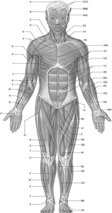
Using the above-referenced diagrammatic anterior view of the major superficial axial and appendicular muscles, identify the specified labeled item(s)in each of the following questions.
Identify the structure(s) indicated by Label Q.
A) Tensor fasciae latae
B) Sartorius muscle
C) Adductor longus muscle
D) Rectus femoris muscle
E) Gracilis muscle
Question
Figure 10.1
Using the above-referenced diagrammatic anterior view of the major superficial axial and appendicular muscles, identify the specified labeled item(s)in each of the following questions.
Identify the structure(s) indicated by Label Y.
A) Calcaneal tendon
B) Linea alba
C) Flexor retinaculum
D) Superior extensor retinaculum
E) Rectus sheath
Question
Figure 10.1
Using the above-referenced diagrammatic anterior view of the major superficial axial and appendicular muscles, identify the specified labeled item(s)in each of the following questions.
Identify the structure(s) indicated by Label P.
A) Flexor retinaculum
B) Iliotibial tract
C) Extensor retinaculum
D) Linea alba
E) Rectus sheath
Question
Figure 10.1
Using the above-referenced diagrammatic anterior view of the major superficial axial and appendicular muscles, identify the specified labeled item(s)in each of the following questions.
Identify the structure(s) indicated by Label T.
A) Gracilis
B) Tensor fasciae latae
C) Iliotibial tract
D) Sartorius
E) Fibularis longus
Question
Figure 10.1
Using the above-referenced diagrammatic anterior view of the major superficial axial and appendicular muscles, identify the specified labeled item(s)in each of the following questions.
Identify the structure(s) indicated by Label A.
A) Tendinous inscriptions
B) Epicranial aponeurosis
C) Linea alba
D) Iliotibial tract
E) Superior extensor retinaculum
Question
Figure 10.1
Using the above-referenced diagrammatic anterior view of the major superficial axial and appendicular muscles, identify the specified labeled item(s)in each of the following questions.
Identify the structure(s) indicated by Label G.
A) Biceps brachii muscle
B) Brachialis muscle
C) Triceps brachii muscle
D) Brachioradialis muscle
E) Pronator teres muscle
Question
Figure 10.1
Using the above-referenced diagrammatic anterior view of the major superficial axial and appendicular muscles, identify the specified labeled item(s)in each of the following questions.
Identify the structure(s) indicated by Label W.
A) Extensor digitorum longus
B) Soleus muscle
C) Gastrocnemius muscle
D) Fibularis longus
E) Tibialis anterior
Question
Question
Figure 10.1
Using the above-referenced diagrammatic anterior view of the major superficial axial and appendicular muscles, identify the specified labeled item(s)in each of the following questions.
Identify the structure(s) indicated by Label X.
A) Soleus muscle
B) Extensor digitorum longus
C) Tibialis anterior
D) Lateral malleolus of fibula
E) Fibularis longus
Question
Figure 10.1
Using the above-referenced diagrammatic anterior view of the major superficial axial and appendicular muscles, identify the specified labeled item(s)in each of the following questions.
Identify the structure(s) indicated by Label R.
A) Vastus lateralis muscle
B) Iliopsoas muscle
C) Pectineus muscle
D) Rectus femoris muscle
E) Adductor longus muscle
Question
Figure 10.1
Using the above-referenced diagrammatic anterior view of the major superficial axial and appendicular muscles, identify the specified labeled item(s)in each of the following questions.
Identify the structure(s) indicated by Label M.
A) Flexor digitorum superficialis muscle
B) Palmaris longus muscle
C) Brachioradialis muscle
D) Pronator teres muscle
E) Extensor carpi radialis brevis muscle
Question
Figure 10.1
Using the above-referenced diagrammatic anterior view of the major superficial axial and appendicular muscles, identify the specified labeled item(s)in each of the following questions.
Identify the structure(s) indicated by Label F.
A) Serratus anterior muscle
B) Deltoid muscle
C) Pectoralis major muscle
D) External oblique muscle
E) Teres major muscle
Question
Question
Question
Figure 10.1
Using the above-referenced diagrammatic anterior view of the major superficial axial and appendicular muscles, identify the specified labeled item(s)in each of the following questions.
Identify the structure(s) indicated by Label S.
A) Iliotibial tract
B) Vastus lateralis muscle
C) Tensor fasciae latae
D) Fibularis longus muscle
E) Pectineus muscle
Question
Figure 10.1
Using the above-referenced diagrammatic anterior view of the major superficial axial and appendicular muscles, identify the specified labeled item(s)in each of the following questions.
Identify the structure(s) indicated by Label E.
A) Triceps brachii muscle
B) Trapezius muscle
C) Clavicle
D) Pectoralis major muscle
E) Deltoid muscle
Question
Question
Figure 10.1
Using the above-referenced diagrammatic anterior view of the major superficial axial and appendicular muscles, identify the specified labeled item(s)in each of the following questions.
Identify the structure(s) indicated by Label L.
A) Extensor carpi radialis longus muscle
B) Brachioradialis muscle
C) Palmaris longus muscle
D) Flexor carpi radialis muscle
E) Pronator teres muscle
Question
Figure 10.1
Using the above-referenced diagrammatic anterior view of the major superficial axial and appendicular muscles, identify the specified labeled item(s)in each of the following questions.
Identify the structure(s) indicated by Label C.
A) Deltoid muscle
B) Latissimus dorsi muscle
C) Sternocleidomastoid muscle
D) Trapezius muscle
E) Occipitofrontalis muscle
Question
Figure 10.1
Using the above-referenced diagrammatic anterior view of the major superficial axial and appendicular muscles, identify the specified labeled item(s)in each of the following questions.
Identify the structure(s) indicated by Label K.
A) Palmaris longus muscle
B) Pronator teres muscle
C) Brachialis muscle
D) Biceps brachii muscle
E) Brachioradialis muscle

Unlock Deck
Sign up to unlock the cards in this deck!
Unlock Deck
Unlock Deck
1/142
Play
Full screen (f)
Deck 10: The Muscular System: Axial Musculature
1
Axial muscles fall into logical groups based upon ________.
A) muscle fiber pattern
B) muscle shape and function
C) muscle structure
D) location and function
E) None of the answers are correct.
A) muscle fiber pattern
B) muscle shape and function
C) muscle structure
D) location and function
E) None of the answers are correct.
D
2
Muscles of the head and neck that are innervated by the trochlear nerve are associated with ________.
A) those that govern feeding
B) those that govern verbal communication
C) actions that form facial expressions
D) actions that orient the eyes
E) None of the answers are correct.
A) those that govern feeding
B) those that govern verbal communication
C) actions that form facial expressions
D) actions that orient the eyes
E) None of the answers are correct.
D
3
Which of the following belongs to the second group of the axial muscles that include flexors and extensors of the axial skeleton?
A) the muscles of the vertebral column
B) the muscles of the perineum
C) the muscles of the head and neck
D) the oblique and rectus muscles
E) the muscles of the pelvic diaphragm
A) the muscles of the vertebral column
B) the muscles of the perineum
C) the muscles of the head and neck
D) the oblique and rectus muscles
E) the muscles of the pelvic diaphragm
A
4
Which of the following muscles runs between the larynx and the hyoid bone?
A) thyrohyoid
B) digastric
C) mylohyoid
D) genioglossus
E) geniohyoid
A) thyrohyoid
B) digastric
C) mylohyoid
D) genioglossus
E) geniohyoid
Unlock Deck
Unlock for access to all 142 flashcards in this deck.
Unlock Deck
k this deck
5
The ________ pulls the eyebrow skin inferiorly and medially, and wrinkles the brow.
A) orbicularis oculi
B) levator palpebrae superioris
C) procerus
D) levator anguli oris
E) corrugator supercilii
A) orbicularis oculi
B) levator palpebrae superioris
C) procerus
D) levator anguli oris
E) corrugator supercilii
Unlock Deck
Unlock for access to all 142 flashcards in this deck.
Unlock Deck
k this deck
6
Which of the following muscles originates at the mandible bone?
A) thyrohyoid
B) geniohyoid
C) stylohyoid
D) styloglossus
E) None of the answers are correct.
A) thyrohyoid
B) geniohyoid
C) stylohyoid
D) styloglossus
E) None of the answers are correct.
Unlock Deck
Unlock for access to all 142 flashcards in this deck.
Unlock Deck
k this deck
7
Which of the following is/are the major action(s)of the anterior neck musculature?
A) depress the mandible
B) stabilize the muscles of the tongue and pharynx
C) tense the floor of the mouth
D) control the position of the larynx
E) All of the answers are correct.
A) depress the mandible
B) stabilize the muscles of the tongue and pharynx
C) tense the floor of the mouth
D) control the position of the larynx
E) All of the answers are correct.
Unlock Deck
Unlock for access to all 142 flashcards in this deck.
Unlock Deck
k this deck
8
The ________ muscle elevates the larynx and is innervated by cranial nerve VII.
A) sternohyoid
B) thyrohyoid
C) stylohyoid
D) omohyoid
E) mylohyoid
A) sternohyoid
B) thyrohyoid
C) stylohyoid
D) omohyoid
E) mylohyoid
Unlock Deck
Unlock for access to all 142 flashcards in this deck.
Unlock Deck
k this deck
9
Orbicularis oris, orbicularis oculi, and platysma are part of the group of muscles known as the muscles of ________.
A) facial expression
B) mastication
C) the extra-ocular region
D) the tongue
E) the pharynx
A) facial expression
B) mastication
C) the extra-ocular region
D) the tongue
E) the pharynx
Unlock Deck
Unlock for access to all 142 flashcards in this deck.
Unlock Deck
k this deck
10
Muscles of mastication most often insert into the ________.
A) mandible
B) bones of the face
C) tongue
D) muscles adjacent to them
E) eyes
A) mandible
B) bones of the face
C) tongue
D) muscles adjacent to them
E) eyes
Unlock Deck
Unlock for access to all 142 flashcards in this deck.
Unlock Deck
k this deck
11
The trigeminal nerve controls which group of muscles?
A) muscles of facial expression
B) muscles of mastication
C) muscles of the tongue
D) muscles of the pharynx
E) muscles of the eye
A) muscles of facial expression
B) muscles of mastication
C) muscles of the tongue
D) muscles of the pharynx
E) muscles of the eye
Unlock Deck
Unlock for access to all 142 flashcards in this deck.
Unlock Deck
k this deck
12
The muscular system is divided into axial and appendicular divisions. The other system that is similarly divided is the ________ system.
A) integument
B) nervous
C) digestive
D) skeletal
E) circulatory
A) integument
B) nervous
C) digestive
D) skeletal
E) circulatory
Unlock Deck
Unlock for access to all 142 flashcards in this deck.
Unlock Deck
k this deck
13
Which of the following muscles is inferior to the lips?
A) masseter
B) frontalis
C) depressor anguli oris
D) risorius
E) zygomaticus major
A) masseter
B) frontalis
C) depressor anguli oris
D) risorius
E) zygomaticus major
Unlock Deck
Unlock for access to all 142 flashcards in this deck.
Unlock Deck
k this deck
14
Which of the following muscles originates at two heads?
A) sternocleidomastoid
B) sternohyoid
C) thyrohyoid
D) mylohyoid
E) stylohyoid
A) sternocleidomastoid
B) sternohyoid
C) thyrohyoid
D) mylohyoid
E) stylohyoid
Unlock Deck
Unlock for access to all 142 flashcards in this deck.
Unlock Deck
k this deck
15
The facial nerve supplies the muscles of ________.
A) the tongue
B) the eyes
C) mastication
D) the anterior neck
E) facial expression
A) the tongue
B) the eyes
C) mastication
D) the anterior neck
E) facial expression
Unlock Deck
Unlock for access to all 142 flashcards in this deck.
Unlock Deck
k this deck
16
Which of the following muscles is the most powerful muscle involved in the process of chewing or manipulating food in the mouth?
A) masseter
B) omohyoid
C) temporalis
D) pterygoid
E) hyoglossus
A) masseter
B) omohyoid
C) temporalis
D) pterygoid
E) hyoglossus
Unlock Deck
Unlock for access to all 142 flashcards in this deck.
Unlock Deck
k this deck
17
How do the extra-ocular eye muscles differ in action from the intrinsic eye muscles?
A) The two muscle groups do not differ in movement types, only in when the movements occur.
B) The extra-ocular muscles cause faster movements than do the intrinsic muscles.
C) Extra-ocular eye muscles move the eyeball in relation to the rest of the body, whereas intrinsic muscles move structures within the eyeball.
D) The extra-ocular eye muscles are smooth muscles inside the eyeball while the intrinsic eye muscles originate on the surface of the orbit and insert into the sclera of the eye just posterior to the cornea.
E) None of the answers are correct.
Unlock Deck
Unlock for access to all 142 flashcards in this deck.
Unlock Deck
k this deck
18
Which muscle(s)moves the bolus into the esophagus?
A) pharyngeal constrictors
B) styloglossus
C) levator veli palatini
D) salpingopharyngeus
E) palatopharyngeus
A) pharyngeal constrictors
B) styloglossus
C) levator veli palatini
D) salpingopharyngeus
E) palatopharyngeus
Unlock Deck
Unlock for access to all 142 flashcards in this deck.
Unlock Deck
k this deck
19
Which of the following features are common to the muscles of mastication?
A) They share an oculomotor nerve innervation.
B) They move the mandible at the temporomandibular joint.
C) They are primarily grouped among the muscles of facial expression.
D) They allow a person to smile.
E) They control swallowing.
A) They share an oculomotor nerve innervation.
B) They move the mandible at the temporomandibular joint.
C) They are primarily grouped among the muscles of facial expression.
D) They allow a person to smile.
E) They control swallowing.
Unlock Deck
Unlock for access to all 142 flashcards in this deck.
Unlock Deck
k this deck
20
Which of the following muscles has an origin on the temporal bone?
A) palatoglossus muscle
B) hyoglossus muscle
C) styloglossus muscle
D) genioglossus muscle
E) None of the answers are correct.
A) palatoglossus muscle
B) hyoglossus muscle
C) styloglossus muscle
D) genioglossus muscle
E) None of the answers are correct.
Unlock Deck
Unlock for access to all 142 flashcards in this deck.
Unlock Deck
k this deck
21
The oblique muscle and/or rectus muscle do which of the following?
A) rotate the vertebral column
B) compress underlying structures
C) occur in the neck
D) important role in respiratory movements of the ribs
E) All of the answers are correct.
A) rotate the vertebral column
B) compress underlying structures
C) occur in the neck
D) important role in respiratory movements of the ribs
E) All of the answers are correct.
Unlock Deck
Unlock for access to all 142 flashcards in this deck.
Unlock Deck
k this deck
22
The erector spinae muscle group that is located most medial to the vertebral column is the ________.
A) longissimus group
B) spinalis group
C) iliocostalis group
D) capitis group
E) thoracis group
A) longissimus group
B) spinalis group
C) iliocostalis group
D) capitis group
E) thoracis group
Unlock Deck
Unlock for access to all 142 flashcards in this deck.
Unlock Deck
k this deck
23
Which of the following extends the vertebral column and depresses the ribs?
A) longus capitis
B) iliocostalis lumborum
C) longus cervicis
D) quadratus lumborum
E) longus colli
A) longus capitis
B) iliocostalis lumborum
C) longus cervicis
D) quadratus lumborum
E) longus colli
Unlock Deck
Unlock for access to all 142 flashcards in this deck.
Unlock Deck
k this deck
24
The diaphragm muscle is innervated by ________.
A) cranial nerve X
B) the phrenic nerves
C) the intercostal nerves
D) the subcostal nerves
E) the thoracic nerves
A) cranial nerve X
B) the phrenic nerves
C) the intercostal nerves
D) the subcostal nerves
E) the thoracic nerves
Unlock Deck
Unlock for access to all 142 flashcards in this deck.
Unlock Deck
k this deck
25
The muscular partition that separates the abdominopelvic and thoracic cavities is the ________.
A) masseter
B) transversus abdominis
C) diaphragm
D) perineum
E) rectus abdominis
A) masseter
B) transversus abdominis
C) diaphragm
D) perineum
E) rectus abdominis
Unlock Deck
Unlock for access to all 142 flashcards in this deck.
Unlock Deck
k this deck
26
The muscle that originates on the sacrum and transverse process of each vertebra and inserts on the spinous process of the third or fourth more superior vertebra is the ________ muscle.
A) quadratus lumborum
B) interspinalis
C) semispinalis
D) multifidus
E) rotatores
A) quadratus lumborum
B) interspinalis
C) semispinalis
D) multifidus
E) rotatores
Unlock Deck
Unlock for access to all 142 flashcards in this deck.
Unlock Deck
k this deck
27
The muscle that assists in mastication, and is also useful to musicians, as in playing a trumpet, is the ________ muscle.
A) masseter
B) orbicularis oris
C) procerus
D) temporalis
E) buccinator
A) masseter
B) orbicularis oris
C) procerus
D) temporalis
E) buccinator
Unlock Deck
Unlock for access to all 142 flashcards in this deck.
Unlock Deck
k this deck
28
Botox is directly injected into the muscles of ________.
A) the tongue
B) facial expression
C) the pharynx
D) the extra-ocular region
E) mastication
A) the tongue
B) facial expression
C) the pharynx
D) the extra-ocular region
E) mastication
Unlock Deck
Unlock for access to all 142 flashcards in this deck.
Unlock Deck
k this deck
29
The digastric and omohyoid muscles are similar in that they both ________.
A) depress the mandible
B) have origins on the inferior surface of the mandible at the chin
C) depress the hyoid bone
D) have two bellies
E) originate from the superior border of the scapula near the suprascapular notch
A) depress the mandible
B) have origins on the inferior surface of the mandible at the chin
C) depress the hyoid bone
D) have two bellies
E) originate from the superior border of the scapula near the suprascapular notch
Unlock Deck
Unlock for access to all 142 flashcards in this deck.
Unlock Deck
k this deck
30
The ________ muscle originates on the horns of the hyoid bone and inserts in the median raphe, and is innervated by branches of the pharyngeal plexus.
A) middle pharyngeal constrictor
B) salpingopharyngeus
C) inferior pharyngeal constrictor
D) palatopharyngeus
E) superior pharyngeal constrictor
A) middle pharyngeal constrictor
B) salpingopharyngeus
C) inferior pharyngeal constrictor
D) palatopharyngeus
E) superior pharyngeal constrictor
Unlock Deck
Unlock for access to all 142 flashcards in this deck.
Unlock Deck
k this deck
31
Which of the following can flex the neck?
A) iliocostalis lumborum
B) iliocostalis cervicis
C) longissimus thoracis
D) longus colli
E) multifidus
A) iliocostalis lumborum
B) iliocostalis cervicis
C) longissimus thoracis
D) longus colli
E) multifidus
Unlock Deck
Unlock for access to all 142 flashcards in this deck.
Unlock Deck
k this deck
32
The intermediate layer of the intrinsic back muscles is also called the ________ group.
A) erector spinae
B) spinal flexor
C) intertransversarii
D) rotatores
E) interspinalis
A) erector spinae
B) spinal flexor
C) intertransversarii
D) rotatores
E) interspinalis
Unlock Deck
Unlock for access to all 142 flashcards in this deck.
Unlock Deck
k this deck
33
The ________ muscle is divided longitudinally by a median collagenous partition called the linea alba.
A) rectus abdominis
B) internal oblique
C) transversus abdominis
D) diaphragm
E) external oblique
A) rectus abdominis
B) internal oblique
C) transversus abdominis
D) diaphragm
E) external oblique
Unlock Deck
Unlock for access to all 142 flashcards in this deck.
Unlock Deck
k this deck
34
The muscle that originates on the lateral nasal cartilages and the aponeuroses covering the inferior portions of the nasal bones, and draws the medial angle of eyebrows inferiorly is called the ________.
A) mentalis
B) procerus
C) occipitofrontalis
D) nasalis
E) corrugator supercilii
A) mentalis
B) procerus
C) occipitofrontalis
D) nasalis
E) corrugator supercilii
Unlock Deck
Unlock for access to all 142 flashcards in this deck.
Unlock Deck
k this deck
35
Of the following muscles, which can compress the abdomen?
A) internal oblique
B) external oblique
C) transversus abdominis
D) rectus abdominis
E) All of the answers are correct.
A) internal oblique
B) external oblique
C) transversus abdominis
D) rectus abdominis
E) All of the answers are correct.
Unlock Deck
Unlock for access to all 142 flashcards in this deck.
Unlock Deck
k this deck
36
The deepest lateral layer of the abdominal muscles is the ________.
A) transversus abdominis
B) rectus abdominis
C) internal oblique
D) external oblique
E) psoas major
A) transversus abdominis
B) rectus abdominis
C) internal oblique
D) external oblique
E) psoas major
Unlock Deck
Unlock for access to all 142 flashcards in this deck.
Unlock Deck
k this deck
37
A muscle that elevates the corner of the mouth and draws it laterally is the ________ muscle.
A) levator anguli oris
B) zygomaticus major
C) risorius
D) depressor anguli oris
E) orbicularis oris
A) levator anguli oris
B) zygomaticus major
C) risorius
D) depressor anguli oris
E) orbicularis oris
Unlock Deck
Unlock for access to all 142 flashcards in this deck.
Unlock Deck
k this deck
38
Which of the following is the function of the superficial layer of the intrinsic back muscles?
A) flexion of the vertebral column
B) extension or lateral flexion of the neck
C) interconnect and stabilize the vertebrae
D) hyperextension of the vertebral column
E) depress the ribs
A) flexion of the vertebral column
B) extension or lateral flexion of the neck
C) interconnect and stabilize the vertebrae
D) hyperextension of the vertebral column
E) depress the ribs
Unlock Deck
Unlock for access to all 142 flashcards in this deck.
Unlock Deck
k this deck
39
These posterior neck muscles extend, rotate, and laterally flex the cervical vertebrae.
A) Iliocostalis group
B) Splenius group
C) Spinalis group
D) Longissimus group
E) None of the answers are correct.
A) Iliocostalis group
B) Splenius group
C) Spinalis group
D) Longissimus group
E) None of the answers are correct.
Unlock Deck
Unlock for access to all 142 flashcards in this deck.
Unlock Deck
k this deck
40
When the ________ muscle contracts, the eye rolls and looks up and laterally.
A) inferior rectus
B) superior oblique
C) inferior oblique
D) superior rectus
E) lateral rectus
A) inferior rectus
B) superior oblique
C) inferior oblique
D) superior rectus
E) lateral rectus
Unlock Deck
Unlock for access to all 142 flashcards in this deck.
Unlock Deck
k this deck
41
Flexion of the vertebral column and depression of the ribs are actions accomplished by the ________ muscles in the abdominal wall.
A) inferior serratus posterior
B) diaphragm
C) rectus abdominis
D) internal intercostals
E) transversus abdominis
A) inferior serratus posterior
B) diaphragm
C) rectus abdominis
D) internal intercostals
E) transversus abdominis
Unlock Deck
Unlock for access to all 142 flashcards in this deck.
Unlock Deck
k this deck
42
The anterior, middle, and posterior scalene muscles are the oblique muscles of the neck, which ________.
A) elevate the first two ribs and/or flex the neck
B) depress the first two ribs and flex the neck
C) elevate the first two ribs and enlarge the thoracic cavity
D) depress the first two ribs and flex the vertebral column
E) elevate the first two ribs and oppose the diaphragm
A) elevate the first two ribs and/or flex the neck
B) depress the first two ribs and flex the neck
C) elevate the first two ribs and enlarge the thoracic cavity
D) depress the first two ribs and flex the vertebral column
E) elevate the first two ribs and oppose the diaphragm
Unlock Deck
Unlock for access to all 142 flashcards in this deck.
Unlock Deck
k this deck
43
In infants, a muscle, which is responsible for producing the suction required for suckling at the breast, is the buccinator.
Unlock Deck
Unlock for access to all 142 flashcards in this deck.
Unlock Deck
k this deck
44
Temporalis is a muscle of facial expression, which moves the auricle of the ear.
Unlock Deck
Unlock for access to all 142 flashcards in this deck.
Unlock Deck
k this deck
45
What would be the consequences of a lacerated trigeminal nerve in terms of muscular control?
Unlock Deck
Unlock for access to all 142 flashcards in this deck.
Unlock Deck
k this deck
46
Tensor veli palatini is a pharyngeal muscle, which elevates the soft palate.
Unlock Deck
Unlock for access to all 142 flashcards in this deck.
Unlock Deck
k this deck
47
A muscle of the scalp, with two bellies separated by a collagenous sheet, is called occipitofrontalis.
Unlock Deck
Unlock for access to all 142 flashcards in this deck.
Unlock Deck
k this deck
48
Which of the following is the most superficial and lateral muscle of the male or female urogenital triangle?
A) ischiocavernosus
B) bulbospongiosus
C) pubococcygeus
D) coccygeus
E) external urethral sphincter
A) ischiocavernosus
B) bulbospongiosus
C) pubococcygeus
D) coccygeus
E) external urethral sphincter
Unlock Deck
Unlock for access to all 142 flashcards in this deck.
Unlock Deck
k this deck
49
Palatoglossus is a muscle of the tongue.
Unlock Deck
Unlock for access to all 142 flashcards in this deck.
Unlock Deck
k this deck
50
The muscle that elevates, everts, and protrudes the lower lip is the mentalis.
Unlock Deck
Unlock for access to all 142 flashcards in this deck.
Unlock Deck
k this deck
51
A superficial muscle that covers the anterior surface of the neck is the trapezius.
Unlock Deck
Unlock for access to all 142 flashcards in this deck.
Unlock Deck
k this deck
52
The deep transverse perineal muscle flexes coccygeal joints, and it elevates and supports the pelvic floor.
Unlock Deck
Unlock for access to all 142 flashcards in this deck.
Unlock Deck
k this deck
53
The internal intercostal muscles aid in respiration by depressing the ribs.
Unlock Deck
Unlock for access to all 142 flashcards in this deck.
Unlock Deck
k this deck
54
A muscle that constricts the opening of the mouth is orbicularis oculi.
Unlock Deck
Unlock for access to all 142 flashcards in this deck.
Unlock Deck
k this deck
55
The superior serratus posterior muscle originates on the spinous processes of C7-T3 and the ligamentum nuchae, and functions in elevating the ribs and enlarging the thoracic cavity.
Unlock Deck
Unlock for access to all 142 flashcards in this deck.
Unlock Deck
k this deck
56
The digastric muscle depresses the mandible and/or elevates the larynx.
Unlock Deck
Unlock for access to all 142 flashcards in this deck.
Unlock Deck
k this deck
57
The risorius is a muscle of mastication.
Unlock Deck
Unlock for access to all 142 flashcards in this deck.
Unlock Deck
k this deck
58
The pubococcygeus muscle, which originates on the inner margins of the pubis and inserts on the coccyx and median raphe, supports the pelvic organs and elevates and retracts the anus.
Unlock Deck
Unlock for access to all 142 flashcards in this deck.
Unlock Deck
k this deck
59
The ________ muscle ejects urine or semen in the male.
A) deep transverse perineal
B) superficial transverse perineal
C) ischiocavernosus
D) external urethral sphincter
E) bulbospongiosus
A) deep transverse perineal
B) superficial transverse perineal
C) ischiocavernosus
D) external urethral sphincter
E) bulbospongiosus
Unlock Deck
Unlock for access to all 142 flashcards in this deck.
Unlock Deck
k this deck
60
The external anal sphincter muscle and the pubococcygeus muscle are located within this perineal region.
A) urogenital triangle
B) anal triangle
C) urogenital diaphragm
D) diaphragm muscle
E) both urogenital triangle and anal triangle
A) urogenital triangle
B) anal triangle
C) urogenital diaphragm
D) diaphragm muscle
E) both urogenital triangle and anal triangle
Unlock Deck
Unlock for access to all 142 flashcards in this deck.
Unlock Deck
k this deck
61
Figure 10.1
Using the above-referenced diagrammatic anterior view of the major superficial axial and appendicular muscles, identify the specified labeled item(s)in each of the following questions.
Identify the structure(s) indicated by Label Q.
A) Tensor fasciae latae
B) Sartorius muscle
C) Adductor longus muscle
D) Rectus femoris muscle
E) Gracilis muscle
Unlock Deck
Unlock for access to all 142 flashcards in this deck.
Unlock Deck
k this deck
62
Figure 10.1
Using the above-referenced diagrammatic anterior view of the major superficial axial and appendicular muscles, identify the specified labeled item(s)in each of the following questions.
Identify the structure(s) indicated by Label Y.
A) Calcaneal tendon
B) Linea alba
C) Flexor retinaculum
D) Superior extensor retinaculum
E) Rectus sheath
Unlock Deck
Unlock for access to all 142 flashcards in this deck.
Unlock Deck
k this deck
63
Figure 10.1
Using the above-referenced diagrammatic anterior view of the major superficial axial and appendicular muscles, identify the specified labeled item(s)in each of the following questions.
Identify the structure(s) indicated by Label P.
A) Flexor retinaculum
B) Iliotibial tract
C) Extensor retinaculum
D) Linea alba
E) Rectus sheath
Unlock Deck
Unlock for access to all 142 flashcards in this deck.
Unlock Deck
k this deck
64
Figure 10.1
Using the above-referenced diagrammatic anterior view of the major superficial axial and appendicular muscles, identify the specified labeled item(s)in each of the following questions.
Identify the structure(s) indicated by Label T.
A) Gracilis
B) Tensor fasciae latae
C) Iliotibial tract
D) Sartorius
E) Fibularis longus
Unlock Deck
Unlock for access to all 142 flashcards in this deck.
Unlock Deck
k this deck
65
Figure 10.1
Using the above-referenced diagrammatic anterior view of the major superficial axial and appendicular muscles, identify the specified labeled item(s)in each of the following questions.
Identify the structure(s) indicated by Label A.
A) Tendinous inscriptions
B) Epicranial aponeurosis
C) Linea alba
D) Iliotibial tract
E) Superior extensor retinaculum
Unlock Deck
Unlock for access to all 142 flashcards in this deck.
Unlock Deck
k this deck
66
Figure 10.1
Using the above-referenced diagrammatic anterior view of the major superficial axial and appendicular muscles, identify the specified labeled item(s)in each of the following questions.
Identify the structure(s) indicated by Label G.
A) Biceps brachii muscle
B) Brachialis muscle
C) Triceps brachii muscle
D) Brachioradialis muscle
E) Pronator teres muscle
Unlock Deck
Unlock for access to all 142 flashcards in this deck.
Unlock Deck
k this deck
67
Figure 10.1
Using the above-referenced diagrammatic anterior view of the major superficial axial and appendicular muscles, identify the specified labeled item(s)in each of the following questions.
Identify the structure(s) indicated by Label W.
A) Extensor digitorum longus
B) Soleus muscle
C) Gastrocnemius muscle
D) Fibularis longus
E) Tibialis anterior
Unlock Deck
Unlock for access to all 142 flashcards in this deck.
Unlock Deck
k this deck
68
Why can one clear the ears by swallowing?
Unlock Deck
Unlock for access to all 142 flashcards in this deck.
Unlock Deck
k this deck
69
Figure 10.1
Using the above-referenced diagrammatic anterior view of the major superficial axial and appendicular muscles, identify the specified labeled item(s)in each of the following questions.
Identify the structure(s) indicated by Label X.
A) Soleus muscle
B) Extensor digitorum longus
C) Tibialis anterior
D) Lateral malleolus of fibula
E) Fibularis longus
Unlock Deck
Unlock for access to all 142 flashcards in this deck.
Unlock Deck
k this deck
70
Figure 10.1
Using the above-referenced diagrammatic anterior view of the major superficial axial and appendicular muscles, identify the specified labeled item(s)in each of the following questions.
Identify the structure(s) indicated by Label R.
A) Vastus lateralis muscle
B) Iliopsoas muscle
C) Pectineus muscle
D) Rectus femoris muscle
E) Adductor longus muscle
Unlock Deck
Unlock for access to all 142 flashcards in this deck.
Unlock Deck
k this deck
71
Figure 10.1
Using the above-referenced diagrammatic anterior view of the major superficial axial and appendicular muscles, identify the specified labeled item(s)in each of the following questions.
Identify the structure(s) indicated by Label M.
A) Flexor digitorum superficialis muscle
B) Palmaris longus muscle
C) Brachioradialis muscle
D) Pronator teres muscle
E) Extensor carpi radialis brevis muscle
Unlock Deck
Unlock for access to all 142 flashcards in this deck.
Unlock Deck
k this deck
72
Figure 10.1
Using the above-referenced diagrammatic anterior view of the major superficial axial and appendicular muscles, identify the specified labeled item(s)in each of the following questions.
Identify the structure(s) indicated by Label F.
A) Serratus anterior muscle
B) Deltoid muscle
C) Pectoralis major muscle
D) External oblique muscle
E) Teres major muscle
Unlock Deck
Unlock for access to all 142 flashcards in this deck.
Unlock Deck
k this deck
73
What muscular changes are involved in the breathing process?
Unlock Deck
Unlock for access to all 142 flashcards in this deck.
Unlock Deck
k this deck
74
Why are there so few spinal flexors associated with the anterior surface of the vertebral column, whereas there are a large number of different extensor groups?
Unlock Deck
Unlock for access to all 142 flashcards in this deck.
Unlock Deck
k this deck
75
Figure 10.1
Using the above-referenced diagrammatic anterior view of the major superficial axial and appendicular muscles, identify the specified labeled item(s)in each of the following questions.
Identify the structure(s) indicated by Label S.
A) Iliotibial tract
B) Vastus lateralis muscle
C) Tensor fasciae latae
D) Fibularis longus muscle
E) Pectineus muscle
Unlock Deck
Unlock for access to all 142 flashcards in this deck.
Unlock Deck
k this deck
76
Figure 10.1
Using the above-referenced diagrammatic anterior view of the major superficial axial and appendicular muscles, identify the specified labeled item(s)in each of the following questions.
Identify the structure(s) indicated by Label E.
A) Triceps brachii muscle
B) Trapezius muscle
C) Clavicle
D) Pectoralis major muscle
E) Deltoid muscle
Unlock Deck
Unlock for access to all 142 flashcards in this deck.
Unlock Deck
k this deck
77
What are the muscles of the urogenital triangle, and how do they control the functions of this area?
Unlock Deck
Unlock for access to all 142 flashcards in this deck.
Unlock Deck
k this deck
78
Figure 10.1
Using the above-referenced diagrammatic anterior view of the major superficial axial and appendicular muscles, identify the specified labeled item(s)in each of the following questions.
Identify the structure(s) indicated by Label L.
A) Extensor carpi radialis longus muscle
B) Brachioradialis muscle
C) Palmaris longus muscle
D) Flexor carpi radialis muscle
E) Pronator teres muscle
Unlock Deck
Unlock for access to all 142 flashcards in this deck.
Unlock Deck
k this deck
79
Figure 10.1
Using the above-referenced diagrammatic anterior view of the major superficial axial and appendicular muscles, identify the specified labeled item(s)in each of the following questions.
Identify the structure(s) indicated by Label C.
A) Deltoid muscle
B) Latissimus dorsi muscle
C) Sternocleidomastoid muscle
D) Trapezius muscle
E) Occipitofrontalis muscle
Unlock Deck
Unlock for access to all 142 flashcards in this deck.
Unlock Deck
k this deck
80
Figure 10.1
Using the above-referenced diagrammatic anterior view of the major superficial axial and appendicular muscles, identify the specified labeled item(s)in each of the following questions.
Identify the structure(s) indicated by Label K.
A) Palmaris longus muscle
B) Pronator teres muscle
C) Brachialis muscle
D) Biceps brachii muscle
E) Brachioradialis muscle
Unlock Deck
Unlock for access to all 142 flashcards in this deck.
Unlock Deck
k this deck
Unlock Deck
Unlock for access to all 142 flashcards in this deck.


