Deck 8: Abdomen
Full screen (f)
Question
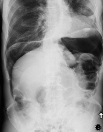 This image shows a patient who:
A) needs a colonoscopy for diagnosis.
B) has a perforated viscus.
C) has a cirrhotic liver.
D) can be treated with IV antibiotics.
Question
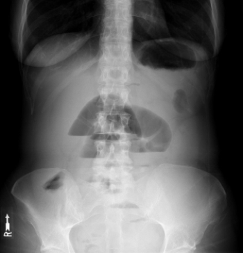 The patient in this image suffers from:
A) a small bowel obstruction.
B) a large bowel obstruction.
C) free intraperitoneal air.
D) appendicitis.
Question
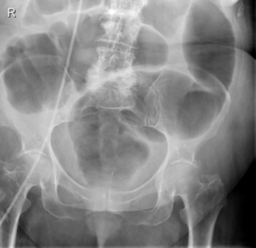 There is an obstruction in this patient's:
A) mid-small bowel.
B) rectum.
C) mid colon.
D) ileocecal valve.
Question
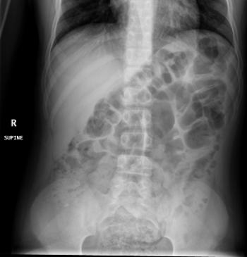 There is a very small but important finding in this patient with acute abdominal pain: What is it?
A) A calcified gallstone
B) Large bowel obstruction
C) Appendicolith
D) Free intraperitoneal air
Question
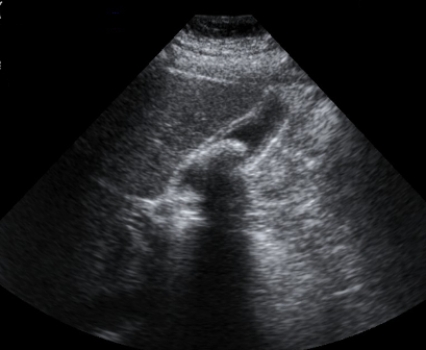 This ultrasound image shows that this patient has:
A) liver cancer.
B) cholelithiasis.
C) liver cyst.
D) blood in Morrison's (hepatorenal) pouch.
Question
Question
Question
Question
Question
Question
Question
Question
Question
Question
Question
Question
Question
Question
Question
Question

Unlock Deck
Sign up to unlock the cards in this deck!
Unlock Deck
Unlock Deck
1/21
Play
Full screen (f)
Deck 8: Abdomen
1
This image shows a patient who:A) needs a colonoscopy for diagnosis.
B) has a perforated viscus.
C) has a cirrhotic liver.
D) can be treated with IV antibiotics.
has a perforated viscus.
2
The patient in this image suffers from:A) a small bowel obstruction.
B) a large bowel obstruction.
C) free intraperitoneal air.
D) appendicitis.
a small bowel obstruction.
3
There is an obstruction in this patient's:A) mid-small bowel.
B) rectum.
C) mid colon.
D) ileocecal valve.
rectum.
4
There is a very small but important finding in this patient with acute abdominal pain: What is it?A) A calcified gallstone
B) Large bowel obstruction
C) Appendicolith
D) Free intraperitoneal air
Unlock Deck
Unlock for access to all 21 flashcards in this deck.
Unlock Deck
k this deck
5
This ultrasound image shows that this patient has:A) liver cancer.
B) cholelithiasis.
C) liver cyst.
D) blood in Morrison's (hepatorenal) pouch.
Unlock Deck
Unlock for access to all 21 flashcards in this deck.
Unlock Deck
k this deck
6
The FAST exam refers to:
A) a rapid protocol CT exam for appendicitis.
B) a very rapid contrast-enhanced radiographic exam for a ruptured aortic aneurysm.
C) an ultrasound exam to detect free fluid in the abdomen.
D) a comprehensive ultrasound evaluation of abdominal and retroperitoneal organs.
A) a rapid protocol CT exam for appendicitis.
B) a very rapid contrast-enhanced radiographic exam for a ruptured aortic aneurysm.
C) an ultrasound exam to detect free fluid in the abdomen.
D) a comprehensive ultrasound evaluation of abdominal and retroperitoneal organs.
Unlock Deck
Unlock for access to all 21 flashcards in this deck.
Unlock Deck
k this deck
7
Which of the following is INCORRECT in the case of acute abdominal pain?
A) Obtain an upright PA chest radiograph.
B) Obtain a supine abdominal AP radiograph.
C) Obtain an upright abdominal radiograph.
D) Obtain a lateral abdominal radiograph.
A) Obtain an upright PA chest radiograph.
B) Obtain a supine abdominal AP radiograph.
C) Obtain an upright abdominal radiograph.
D) Obtain a lateral abdominal radiograph.
Unlock Deck
Unlock for access to all 21 flashcards in this deck.
Unlock Deck
k this deck
8
In a pediatric patient with right lower quadrant pain, your initial imaging modality would likely be:
A) ultrasound.
B) radiography.
C) CT.
D) HIDA scan.
A) ultrasound.
B) radiography.
C) CT.
D) HIDA scan.
Unlock Deck
Unlock for access to all 21 flashcards in this deck.
Unlock Deck
k this deck
9
Ultrasound is never useful for the diagnosis of which of the following conditions?
A) Small bowel obstruction
B) Appendicitis
C) Cholelithiasis
D) Aortic aneurysm
A) Small bowel obstruction
B) Appendicitis
C) Cholelithiasis
D) Aortic aneurysm
Unlock Deck
Unlock for access to all 21 flashcards in this deck.
Unlock Deck
k this deck
10
An abdominal CT scan for a clinically suspected abdominal mass is usually done:
A) using unenhanced scan without IV or oral contrast material.
B) with oral contrast material and without and with IV contrast enhancement.
C) with oral and IV contrast enhancement.
D) with oral contrast material but without IV contrast enhancement.
A) using unenhanced scan without IV or oral contrast material.
B) with oral contrast material and without and with IV contrast enhancement.
C) with oral and IV contrast enhancement.
D) with oral contrast material but without IV contrast enhancement.
Unlock Deck
Unlock for access to all 21 flashcards in this deck.
Unlock Deck
k this deck
11
An abdominal CT scan done to evaluate a hepatic mass seen on ultrasonography:
A) is unlikely to provide a more definitive diagnosis than sonography.
B) should always be done instead of MRI for hepatic mass characterization.
C) is a multiphase study without and with IV contrast enhancement and with delayed scanning.
D) must be done with the injection protocol for CT arteriography.
Unlock Deck
Unlock for access to all 21 flashcards in this deck.
Unlock Deck
k this deck
12
The finding of diffuse increased liver echogenicity on ultrasound (compared to normal) and poor transmission of the ultrasound beam through the liver would suggest:
A) cirrhosis.
B) steatosis.
C) choledocholithiasis.
D) portal hypertension.
A) cirrhosis.
B) steatosis.
C) choledocholithiasis.
D) portal hypertension.
Unlock Deck
Unlock for access to all 21 flashcards in this deck.
Unlock Deck
k this deck
13
Which of the following statements is NOT true of appendicitis?
A) CT is highly reliable in excluding the presence of appendicitis.
B) A conspicuously thickened appendiceal wall and fluid surrounding the appendix on ultrasonography support the diagnosis.
C) An appendicolith may be visible on CT and supports the diagnosis.
D) A decrease in the CT density of fat adjacent to the appendix on CT is supportive of a diagnosis of appendicitis.
Unlock Deck
Unlock for access to all 21 flashcards in this deck.
Unlock Deck
k this deck
14
Which of the following statements IS NOT consistent with diverticulitis?
A) It is typically identified with ultrasonography.
B) It most commonly occurs in the sigmoid colon.
C) It is associated with an increase in the CT density of surrounding fat.
D) In severe cases it may result in abscess formation.
A) It is typically identified with ultrasonography.
B) It most commonly occurs in the sigmoid colon.
C) It is associated with an increase in the CT density of surrounding fat.
D) In severe cases it may result in abscess formation.
Unlock Deck
Unlock for access to all 21 flashcards in this deck.
Unlock Deck
k this deck
15
Which of the following statements pertaining to mesenteric ischemia is INCORRECT?
A) It is associated with the radiographic appearance of "thumbprinting."
B) In severe cases, it can be associated with intramural pneumotosis.
C) It is associated with stenosis in the portal vein.
D) It may result in bowel infarction.
A) It is associated with the radiographic appearance of "thumbprinting."
B) In severe cases, it can be associated with intramural pneumotosis.
C) It is associated with stenosis in the portal vein.
D) It may result in bowel infarction.
Unlock Deck
Unlock for access to all 21 flashcards in this deck.
Unlock Deck
k this deck
16
Which statement is INCORRECT regarding using imaging for patients with suspected jaundice?
A) For clinically suspected obstructive jaundice, the initial imaging procedure is commonly ultrasonography.
B) When a patient presents with obstructive jaundice, weight loss, and vague back pain, MRCP is sufficient for diagnosis.
C) MRCP is used to provide excellent depiction of the biliary and pancreatic ducts.
D) The suspicion that obstruction of the common bile duct caused by a calculus is increased if the patient has many tiny calculi ("biliary gravel") in the gallbladder that are apparent on ultrasound.
Unlock Deck
Unlock for access to all 21 flashcards in this deck.
Unlock Deck
k this deck
17
Which of the following statements regarding GI bleeding is INCORRECT?
A) Ultrasonography may show extravasation into the lumen of a part of the GI tract.
B) CT angiography may show extravasation into the lumen of a part of the GI tract.
C) Catheter angiography may show extravasation into the lumen of a part of the GI tract.
D) In stable patients with relatively slow GI bleeding, a Tc-99m-labeled RBC radionuclide bleeding study may be diagnostic.
Unlock Deck
Unlock for access to all 21 flashcards in this deck.
Unlock Deck
k this deck
18
"Virtual colonoscopy" refers to:
A) a barium enema colonoscopy.
B) CT colonography.
C) MR colonoscopy.
D) colonoscopy using rectal probe ultrasound.
A) a barium enema colonoscopy.
B) CT colonography.
C) MR colonoscopy.
D) colonoscopy using rectal probe ultrasound.
Unlock Deck
Unlock for access to all 21 flashcards in this deck.
Unlock Deck
k this deck
19
Which of the following is NOT true of adrenal masses?
A) Enlargement noted on a routine abdominal CT likely means malignancy.
B) They may be a functional adenoma causing hormonal issues.
C) They may be a pheochromocytoma.
D) Unenhanced CT can reliably identify benign adrenal adenomas.
A) Enlargement noted on a routine abdominal CT likely means malignancy.
B) They may be a functional adenoma causing hormonal issues.
C) They may be a pheochromocytoma.
D) Unenhanced CT can reliably identify benign adrenal adenomas.
Unlock Deck
Unlock for access to all 21 flashcards in this deck.
Unlock Deck
k this deck
20
Following the identification of polyps on a virtual colonoscopy, the radiologist:
A) will likely recommend removal of all polyps.
B) will likely recommend 5-year follow-up exam for all polyps smaller than 5 mm.
C) will likely be more concerned about polyps that are smooth than those that are irregular in shape.
D) can use the American College of Radiology criteria to determine with 99% accuracy whether a polyp needs removal.
Unlock Deck
Unlock for access to all 21 flashcards in this deck.
Unlock Deck
k this deck
21
Which of the following statements regarding liver imaging is INCORRECT?
A) Normally the liver and spleen have close to the same CT density.
B) In hepatic steatosis the liver has a reduced CT density.
C) In liver fibrosis, the liver has an increased CT density.
D) MRI cannot be used to diagnosis liver steatosis.
A) Normally the liver and spleen have close to the same CT density.
B) In hepatic steatosis the liver has a reduced CT density.
C) In liver fibrosis, the liver has an increased CT density.
D) MRI cannot be used to diagnosis liver steatosis.
Unlock Deck
Unlock for access to all 21 flashcards in this deck.
Unlock Deck
k this deck
Unlock Deck
Unlock for access to all 21 flashcards in this deck.


