Deck 4: Brain
Full screen (f)
Question
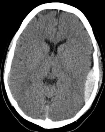 This image reveals the presence of:
A) an epidural hematoma.
B) a subdural hematoma.
C) an acute ischemic stroke.
D) subarachnoid hemorrhage.
Question
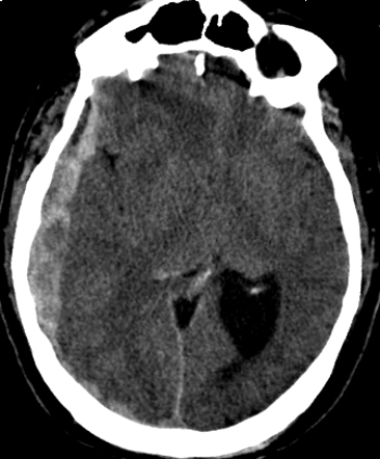 The patient in this image has which of these conditions?
A) An epidural hematoma
B) A subdural hematoma
C) An acute ischemic stroke
D) A subarachnoid hemorrhage
Question
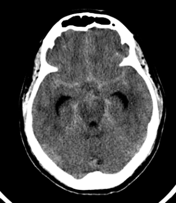 The pathology shown in the image above is associated with:
A) chronic headache.
B) acute onset of aphasia.
C) chronic left-sided weakness.
D) acute onset of what was likely reported as the "worst headache of my life."
Question
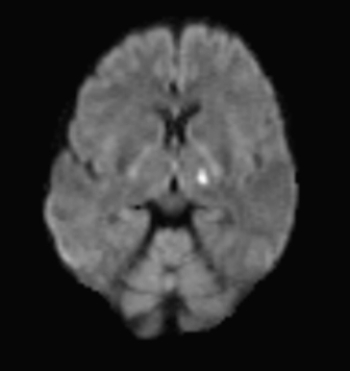 The very bright intensity in the above DWI indicates that this patient:
A) has metastatic disease.
B) had an acute ischemic stroke.
C) has an AVM.
D) has MS.
Question
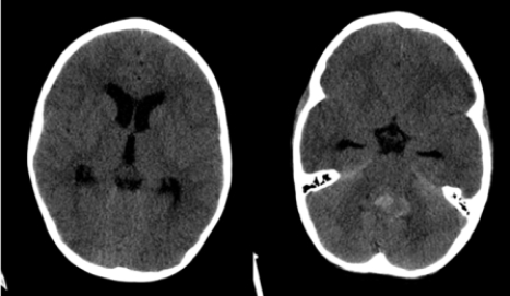 The axial CT images shown are two different slices from one study on a pediatric patient. Your likely diagnosis is:
A) hemorrhagic stroke.
B) obstructive hydrocephalus.
C) subarachnoid hemorrhage.
D) uncal herniation.
Question
 The patient in this image has:
A) a meningioma.
B) hydrocephalus.
C) a cephalhematoma.
D) uncal herniation.
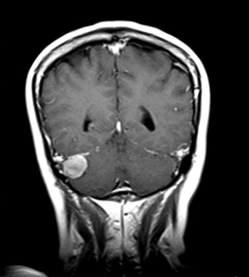
Question
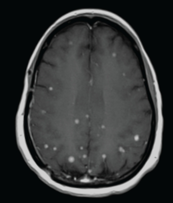 The above axial contrast-enhanced MRI is consistent with:
A) dementia.
B) metastatic breast cancer.
C) Kuru.
D) TIAs.
Question
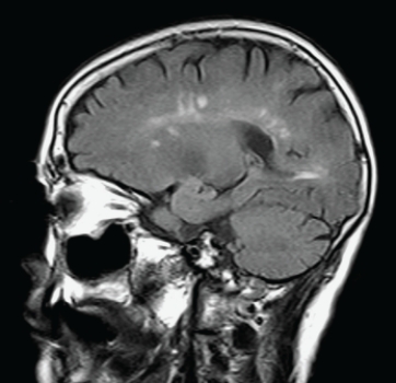 This sagittal T2 FLAIR MRI is consistent with:
A) MS.
B) ischemic stroke.
C) primary brain tumor.
D) pituitary adenoma.
Question
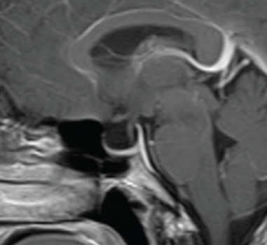 This sagittal contrast-enhanced T1 MRI shows a patient with:
A) visual field defects.
B) chronic headache and papilledema.
C) lower extremity weakness.
D) facial paresthesias.
Question
Question
Question
Question
Question
Question
Question
Question
Question
Question
Question

Unlock Deck
Sign up to unlock the cards in this deck!
Unlock Deck
Unlock Deck
1/20
Play
Full screen (f)
Deck 4: Brain
1
This image reveals the presence of:A) an epidural hematoma.
B) a subdural hematoma.
C) an acute ischemic stroke.
D) subarachnoid hemorrhage.
an epidural hematoma.
2
The patient in this image has which of these conditions?A) An epidural hematoma
B) A subdural hematoma
C) An acute ischemic stroke
D) A subarachnoid hemorrhage
A subdural hematoma
3
The pathology shown in the image above is associated with:A) chronic headache.
B) acute onset of aphasia.
C) chronic left-sided weakness.
D) acute onset of what was likely reported as the "worst headache of my life."
acute onset of what was likely reported as the "worst headache of my life."
4
The very bright intensity in the above DWI indicates that this patient:A) has metastatic disease.
B) had an acute ischemic stroke.
C) has an AVM.
D) has MS.
Unlock Deck
Unlock for access to all 20 flashcards in this deck.
Unlock Deck
k this deck
5
The axial CT images shown are two different slices from one study on a pediatric patient. Your likely diagnosis is:A) hemorrhagic stroke.
B) obstructive hydrocephalus.
C) subarachnoid hemorrhage.
D) uncal herniation.
Unlock Deck
Unlock for access to all 20 flashcards in this deck.
Unlock Deck
k this deck
6
The patient in this image has:A) a meningioma.
B) hydrocephalus.
C) a cephalhematoma.
D) uncal herniation.
Unlock Deck
Unlock for access to all 20 flashcards in this deck.
Unlock Deck
k this deck
7
The above axial contrast-enhanced MRI is consistent with:A) dementia.
B) metastatic breast cancer.
C) Kuru.
D) TIAs.
Unlock Deck
Unlock for access to all 20 flashcards in this deck.
Unlock Deck
k this deck
8
This sagittal T2 FLAIR MRI is consistent with:A) MS.
B) ischemic stroke.
C) primary brain tumor.
D) pituitary adenoma.
Unlock Deck
Unlock for access to all 20 flashcards in this deck.
Unlock Deck
k this deck
9
This sagittal contrast-enhanced T1 MRI shows a patient with:A) visual field defects.
B) chronic headache and papilledema.
C) lower extremity weakness.
D) facial paresthesias.
Unlock Deck
Unlock for access to all 20 flashcards in this deck.
Unlock Deck
k this deck
10
For a patient with closed head trauma for whom imaging is needed, the initial imaging modality of choice is:
A) radiography.
B) unenhanced CT.
C) MRI without and with contrast enhancement.
D) ultrasound.
A) radiography.
B) unenhanced CT.
C) MRI without and with contrast enhancement.
D) ultrasound.
Unlock Deck
Unlock for access to all 20 flashcards in this deck.
Unlock Deck
k this deck
11
There is a narrow therapeutic window for one of your patients with signs of an acute stroke. Which of the following is likely to be done in this narrow window of time?
A) Carotid Doppler ultrasound done at the bedside in the ED with CT arteriography if positive
B) Stat CT scan (and stat MRI if the CT shows no hemorrhage)
C) Cerebral radionuclide PET scan
D) Echocardiography to search for sources of thromboembolic disease to the cerebral circulation
A) Carotid Doppler ultrasound done at the bedside in the ED with CT arteriography if positive
B) Stat CT scan (and stat MRI if the CT shows no hemorrhage)
C) Cerebral radionuclide PET scan
D) Echocardiography to search for sources of thromboembolic disease to the cerebral circulation
Unlock Deck
Unlock for access to all 20 flashcards in this deck.
Unlock Deck
k this deck
12
Diffusion-weighted MRI sequences (DWI) are the BEST modality for detecting which of the following?
A) Acute and early subacute ischemic stroke
B) Hyperacute hemorrhagic stroke
C) MS and other white matter diseases
D) Intracranial venous thrombosis
A) Acute and early subacute ischemic stroke
B) Hyperacute hemorrhagic stroke
C) MS and other white matter diseases
D) Intracranial venous thrombosis
Unlock Deck
Unlock for access to all 20 flashcards in this deck.
Unlock Deck
k this deck
13
A focus of intracranial magnetic susceptibility is associated with:
A) white matter disease.
B) old episode of intracranial bleeding.
C) pineal gland hypertrophy.
D) acute intracranial arterial occlusion.
A) white matter disease.
B) old episode of intracranial bleeding.
C) pineal gland hypertrophy.
D) acute intracranial arterial occlusion.
Unlock Deck
Unlock for access to all 20 flashcards in this deck.
Unlock Deck
k this deck
14
Which of the following would NOT be expected to be seen in an unenhanced CT scan of a patient with an acute cerebral infarction?
A) Insular ribbon sign
B) Hyperdense vessel sign
C) Obscuration of the basal ganglia
D) Dural tail sign
A) Insular ribbon sign
B) Hyperdense vessel sign
C) Obscuration of the basal ganglia
D) Dural tail sign
Unlock Deck
Unlock for access to all 20 flashcards in this deck.
Unlock Deck
k this deck
15
Which of the following statements about vasogenic edema is NOT true?
A) It can be associated with a tumor.
B) It has a positive mass effect.
C) It may result from aging.
D) It appears as increased T2 signal in MRI.
A) It can be associated with a tumor.
B) It has a positive mass effect.
C) It may result from aging.
D) It appears as increased T2 signal in MRI.
Unlock Deck
Unlock for access to all 20 flashcards in this deck.
Unlock Deck
k this deck
16
For MRI of the brain, the use of gadolinium-based contrast agents is usually avoided in which of the following types of patients?
A) Pregnant
B) Elderly
C) Children
D) Those with heart disease
A) Pregnant
B) Elderly
C) Children
D) Those with heart disease
Unlock Deck
Unlock for access to all 20 flashcards in this deck.
Unlock Deck
k this deck
17
White matter disease is usually evident on:
A) the T2 weighted images used in MRI of the brain.
B) only the contrast-enhanced MRI sequences of the brain.
C) intracranial CT arteriography.
D) cisternography.
A) the T2 weighted images used in MRI of the brain.
B) only the contrast-enhanced MRI sequences of the brain.
C) intracranial CT arteriography.
D) cisternography.
Unlock Deck
Unlock for access to all 20 flashcards in this deck.
Unlock Deck
k this deck
18
Empty sella syndrome is associated with:
A) hypopituitarism.
B) visual disturbances.
C) NPH.
D) pituitary adenoma.
A) hypopituitarism.
B) visual disturbances.
C) NPH.
D) pituitary adenoma.
Unlock Deck
Unlock for access to all 20 flashcards in this deck.
Unlock Deck
k this deck
19
Disproportionately decreased volume of the hippocampus is associated with:
A) tremors.
B) Alzheimer's disease.
C) dysarthria.
D) ataxia.
A) tremors.
B) Alzheimer's disease.
C) dysarthria.
D) ataxia.
Unlock Deck
Unlock for access to all 20 flashcards in this deck.
Unlock Deck
k this deck
20
UBOs are associated with:
A) epilepsy.
B) normal aging.
C) Parkinson's disease.
D) metastatic disease.
A) epilepsy.
B) normal aging.
C) Parkinson's disease.
D) metastatic disease.
Unlock Deck
Unlock for access to all 20 flashcards in this deck.
Unlock Deck
k this deck
Unlock Deck
Unlock for access to all 20 flashcards in this deck.


