Deck 2: Shoulder, Pelvis, and Limbs
Full screen (f)
Question
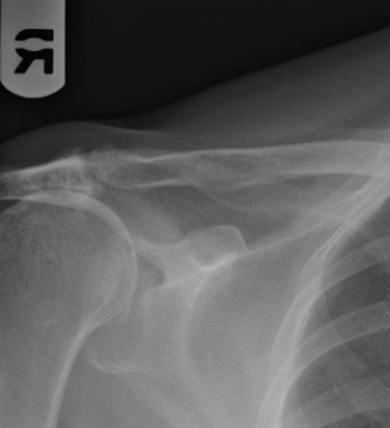
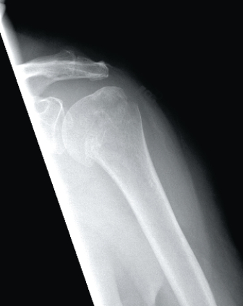 This image depicts a patient with:
A) a Salter-Harris fracture.
B) a normal proximal humerus.
C) an anatomic neck fracture of the humerus.
D) a surgical neck fracture of the humerus.
Question
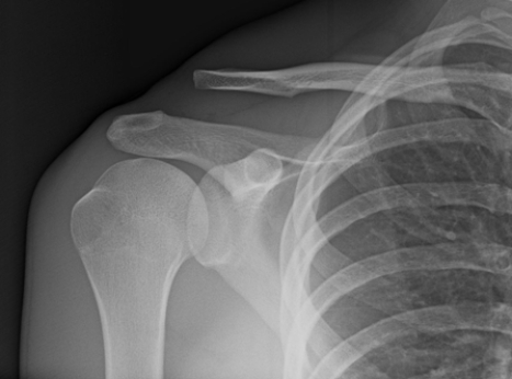
 The patient depicted in the image has:
A) an anterior shoulder dislocation.
B) a normal shoulder in complete adduction.
C) a separated shoulder.
D) a fracture of the proximal humeral shaft.
Question
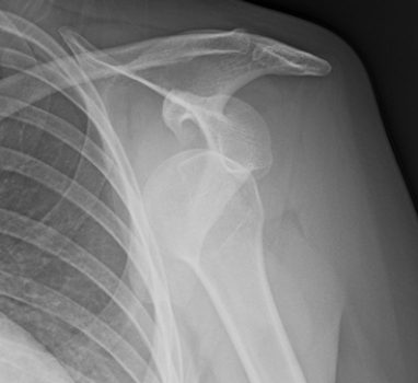
 The patient in this image has:
A) a dislocated shoulder.
B) an AC joint separation.
C) a Bankart lesion.
D) a winged scapula.
Question
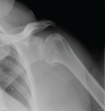 The patient in this image has:
A) glenohumeral joint osteoarthritis.
B) dislocated shoulder.
C) separated shoulder.
D) Bankart lesion.
Question
 This image from a PA chest radiograph shows the incidental finding of:
A) chronic AC joint separation.
B) old nonunited Bankart fracture.
C) a chronic full-thickness supraspinatus tear.
D) blastic metastatic bone lesions.
Question
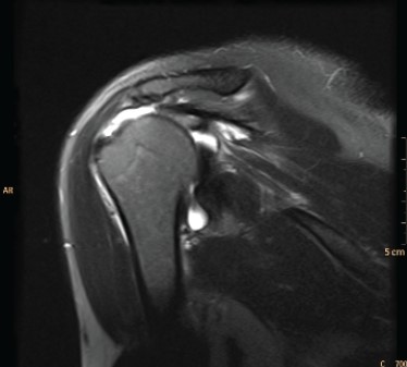 What is the diagnosis based on the coronal T2 MRI?
A) AC joint separation
B) Full-thickness rotator cuff tear
C) Radiographically occult humeral head fracture
D) Glenoid labral tear
Question
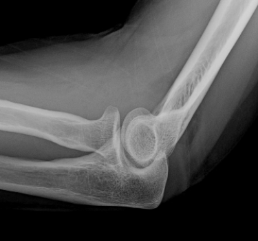 Lateral elbow radiograph after trauma; other radiographs show no fracture. Your diagnosis is:
A) occult intra-articular fracture.
B) dislocated elbow.
C) normal elbow.
D) torn distal biceps tendon tour.
Question
 The patient in this image has:
A) a boxer's fracture.
B) degenerative joint disease of the first carpometacarpal joint.
C) a fractured scaphoid.
D) a spiral second metacarpal fracture.
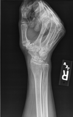
Question
 The image shows a patient with:
A) acute onset of hand pain.
B) a positive rheumatoid arthritis (RA) titer.
C) carpal tunnel syndrome.
D) history of multiple prior hand injuries.
Question
 The patient in this image has:
A) hip joint degenerative joint disease.
B) hip joint dislocation.
C) fractured femoral neck.
D) femoral neck osteophyte.
Question
 Based on the image, what condition/injury does this patient have?
A) A left femoral neck fracture
B) Right hip degenerative joint disease
C) Lytic bone disease
D) Fracture of the right ilium
Question
 The skeletal findings in this patient with a distended bladder (opacified after contrast-enhanced CT) include:
A) right hip dislocation.
B) left pubic rami fractures.
C) right hip joint degenerative joint disease.
D) right femoral neck fracture.
Question
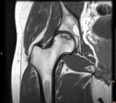 What can you tell about the patient in the MR image?
A) The patient has been on high-dose steroids for a severe rash.
B) The patient is an oncology patient with new onset of hip pain.
C) The patient has hip pain associated with high fever and leukocytosis.
D) The patient has a normal hip; symptoms in the hip are secondary to lumbar disk disease.
Question
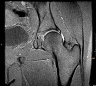 This MR arthrogram depicts a patient with:
A) a normal physical exam of the hip.
B) systemic arthritis.
C) pain and sensation of clicking in the joint.
D) injury suffered while playing shuffleboard.
Question
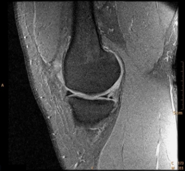
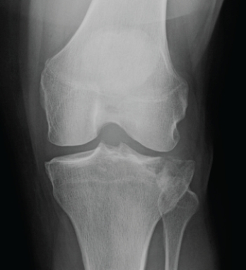
 The patient in this image has:
A) knee joint rheumatoid arthritis.
B) a skiing injury.
C) knee joint osteoarthritis.
D) puncture wound to the knee joint.
Question
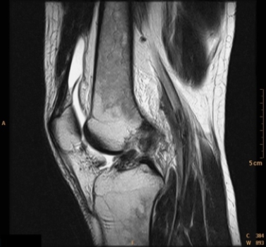
 The MR image shows a patient with a:
A) normal knee.
B) meniscus tear.
C) patella alta.
D) popliteal venous thrombosis.
Question
 Radiographs on this patient, including the cross-table lateral shown here, revealed no visible fracture. How will the patient be managed?
A) Because no fracture is shown, the patient will be told that walking is safe, NSAIDs will be prescribed, and the patient will be referred to physical therapy.
B) Because of the lipohemarthrosis, the patient will be kept on non-weight-bearing and will have an urgent CT or MRI.
C) Radiographic findings above suggest medial meniscus tear. The patient will be scheduled for MRI next week and will be scheduled to see an orthopedic surgeon whenever convenient.
D) Physical examination was unremarkable and the patient was told to go back to work.
Question
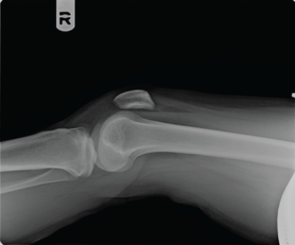
 The patient in this image:
A) has a normal knee.
B) has a Segond fracture.
C) is a patient with Osgood-Schlatter disease.
D) has a tibial plateau fracture.
Question
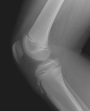 Based on the image of this pediatric patient, what can you tell this patient has?
A) Clinical findings of Osgood-Schlatter disease
B) Acute twisting injury of the knee
C) Tenderness of the quadriceps tendon
D) Popliteal fossa mass
Question
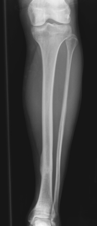 The patient in this image has which of the following injuries/conditions?
A) Tibial osteosarcoma
B) Lateral malleolar fracture
C) A healing tibial stress fracture
D) Fibula osteomyelitis
Question
 The patient in this image has suffered what type of injury?
A) A lateral malleolar fracture; stable ankle mortise
B) A lateral malleolar fracture with widening of the ankle mortise
C) A talar dome osteochondral lesion
D) Stress fracture of the first metatarsal
Question
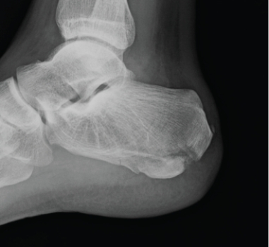 What can you tell about this patient whose lateral calcaneus radiograph is shown?
A) The patient is a long-distance runner with chronic heel pain.
B) The patient has lost the ability to plantar flex at the ankle.
C) The patient has cancer with new onset of foot pain.
D) This patient landed on his feet from a 12-foot fall and also has back pain.
Question
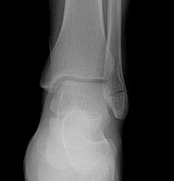
 The patient in the following image has:
A) a normal foot.
B) a Haglund's deformity.
C) a Lisfranc fracture.
D) plantar fasciitis.
Question
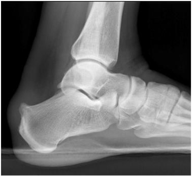
 What specific injury/deformity does the patient in this image suffer from?
A) Rheumatoid arthritis (RA) of the first tarso-metatarsal joint
B) Lisfranc fracture
C) Bunion deformity
D) Haglund's deformity
Question
Question
Question
Question
Question
Question
Question
Question
Question
Question
Question
Question
Question
Question
Question
Question
Question
Question
Question
Question
Question
Question
Question

Unlock Deck
Sign up to unlock the cards in this deck!
Unlock Deck
Unlock Deck
1/47
Play
Full screen (f)
Deck 2: Shoulder, Pelvis, and Limbs
1
This image depicts a patient with:A) a Salter-Harris fracture.
B) a normal proximal humerus.
C) an anatomic neck fracture of the humerus.
D) a surgical neck fracture of the humerus.
an anatomic neck fracture of the humerus.
2
The patient depicted in the image has:A) an anterior shoulder dislocation.
B) a normal shoulder in complete adduction.
C) a separated shoulder.
D) a fracture of the proximal humeral shaft.
an anterior shoulder dislocation.
3
The patient in this image has:A) a dislocated shoulder.
B) an AC joint separation.
C) a Bankart lesion.
D) a winged scapula.
an AC joint separation.
4
The patient in this image has:A) glenohumeral joint osteoarthritis.
B) dislocated shoulder.
C) separated shoulder.
D) Bankart lesion.
Unlock Deck
Unlock for access to all 47 flashcards in this deck.
Unlock Deck
k this deck
5
This image from a PA chest radiograph shows the incidental finding of:A) chronic AC joint separation.
B) old nonunited Bankart fracture.
C) a chronic full-thickness supraspinatus tear.
D) blastic metastatic bone lesions.
Unlock Deck
Unlock for access to all 47 flashcards in this deck.
Unlock Deck
k this deck
6
What is the diagnosis based on the coronal T2 MRI?A) AC joint separation
B) Full-thickness rotator cuff tear
C) Radiographically occult humeral head fracture
D) Glenoid labral tear
Unlock Deck
Unlock for access to all 47 flashcards in this deck.
Unlock Deck
k this deck
7
Lateral elbow radiograph after trauma; other radiographs show no fracture. Your diagnosis is:A) occult intra-articular fracture.
B) dislocated elbow.
C) normal elbow.
D) torn distal biceps tendon tour.
Unlock Deck
Unlock for access to all 47 flashcards in this deck.
Unlock Deck
k this deck
8
The patient in this image has:A) a boxer's fracture.
B) degenerative joint disease of the first carpometacarpal joint.
C) a fractured scaphoid.
D) a spiral second metacarpal fracture.
Unlock Deck
Unlock for access to all 47 flashcards in this deck.
Unlock Deck
k this deck
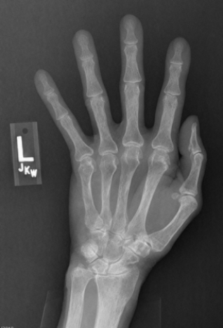
9
The image shows a patient with:A) acute onset of hand pain.
B) a positive rheumatoid arthritis (RA) titer.
C) carpal tunnel syndrome.
D) history of multiple prior hand injuries.
Unlock Deck
Unlock for access to all 47 flashcards in this deck.
Unlock Deck
k this deck
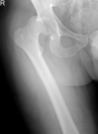
10
The patient in this image has:A) hip joint degenerative joint disease.
B) hip joint dislocation.
C) fractured femoral neck.
D) femoral neck osteophyte.
Unlock Deck
Unlock for access to all 47 flashcards in this deck.
Unlock Deck
k this deck
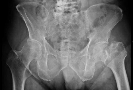
11
Based on the image, what condition/injury does this patient have?A) A left femoral neck fracture
B) Right hip degenerative joint disease
C) Lytic bone disease
D) Fracture of the right ilium
Unlock Deck
Unlock for access to all 47 flashcards in this deck.
Unlock Deck
k this deck
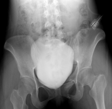
12
The skeletal findings in this patient with a distended bladder (opacified after contrast-enhanced CT) include:A) right hip dislocation.
B) left pubic rami fractures.
C) right hip joint degenerative joint disease.
D) right femoral neck fracture.
Unlock Deck
Unlock for access to all 47 flashcards in this deck.
Unlock Deck
k this deck
13
What can you tell about the patient in the MR image?A) The patient has been on high-dose steroids for a severe rash.
B) The patient is an oncology patient with new onset of hip pain.
C) The patient has hip pain associated with high fever and leukocytosis.
D) The patient has a normal hip; symptoms in the hip are secondary to lumbar disk disease.
Unlock Deck
Unlock for access to all 47 flashcards in this deck.
Unlock Deck
k this deck
14
This MR arthrogram depicts a patient with:A) a normal physical exam of the hip.
B) systemic arthritis.
C) pain and sensation of clicking in the joint.
D) injury suffered while playing shuffleboard.
Unlock Deck
Unlock for access to all 47 flashcards in this deck.
Unlock Deck
k this deck
15
The patient in this image has:A) knee joint rheumatoid arthritis.
B) a skiing injury.
C) knee joint osteoarthritis.
D) puncture wound to the knee joint.
Unlock Deck
Unlock for access to all 47 flashcards in this deck.
Unlock Deck
k this deck
16
The MR image shows a patient with a:A) normal knee.
B) meniscus tear.
C) patella alta.
D) popliteal venous thrombosis.
Unlock Deck
Unlock for access to all 47 flashcards in this deck.
Unlock Deck
k this deck
17
Radiographs on this patient, including the cross-table lateral shown here, revealed no visible fracture. How will the patient be managed?A) Because no fracture is shown, the patient will be told that walking is safe, NSAIDs will be prescribed, and the patient will be referred to physical therapy.
B) Because of the lipohemarthrosis, the patient will be kept on non-weight-bearing and will have an urgent CT or MRI.
C) Radiographic findings above suggest medial meniscus tear. The patient will be scheduled for MRI next week and will be scheduled to see an orthopedic surgeon whenever convenient.
D) Physical examination was unremarkable and the patient was told to go back to work.
Unlock Deck
Unlock for access to all 47 flashcards in this deck.
Unlock Deck
k this deck
18
The patient in this image:A) has a normal knee.
B) has a Segond fracture.
C) is a patient with Osgood-Schlatter disease.
D) has a tibial plateau fracture.
Unlock Deck
Unlock for access to all 47 flashcards in this deck.
Unlock Deck
k this deck
19
Based on the image of this pediatric patient, what can you tell this patient has?A) Clinical findings of Osgood-Schlatter disease
B) Acute twisting injury of the knee
C) Tenderness of the quadriceps tendon
D) Popliteal fossa mass
Unlock Deck
Unlock for access to all 47 flashcards in this deck.
Unlock Deck
k this deck
20
The patient in this image has which of the following injuries/conditions?A) Tibial osteosarcoma
B) Lateral malleolar fracture
C) A healing tibial stress fracture
D) Fibula osteomyelitis
Unlock Deck
Unlock for access to all 47 flashcards in this deck.
Unlock Deck
k this deck
21
The patient in this image has suffered what type of injury?A) A lateral malleolar fracture; stable ankle mortise
B) A lateral malleolar fracture with widening of the ankle mortise
C) A talar dome osteochondral lesion
D) Stress fracture of the first metatarsal
Unlock Deck
Unlock for access to all 47 flashcards in this deck.
Unlock Deck
k this deck
22
What can you tell about this patient whose lateral calcaneus radiograph is shown?A) The patient is a long-distance runner with chronic heel pain.
B) The patient has lost the ability to plantar flex at the ankle.
C) The patient has cancer with new onset of foot pain.
D) This patient landed on his feet from a 12-foot fall and also has back pain.
Unlock Deck
Unlock for access to all 47 flashcards in this deck.
Unlock Deck
k this deck
23
The patient in the following image has:A) a normal foot.
B) a Haglund's deformity.
C) a Lisfranc fracture.
D) plantar fasciitis.
Unlock Deck
Unlock for access to all 47 flashcards in this deck.
Unlock Deck
k this deck
24
What specific injury/deformity does the patient in this image suffer from?A) Rheumatoid arthritis (RA) of the first tarso-metatarsal joint
B) Lisfranc fracture
C) Bunion deformity
D) Haglund's deformity
Unlock Deck
Unlock for access to all 47 flashcards in this deck.
Unlock Deck
k this deck
25
The Salter-Harris classification refers to:
A) the location of fractures associated with growth plates in the immature skeleton.
B) the degree of hip subluxation in hip degenerative joint disorder.
C) the degree of femoral angulation relative to the tibia.
D) the laxity in the pubic bones associated with pregnancy.
A) the location of fractures associated with growth plates in the immature skeleton.
B) the degree of hip subluxation in hip degenerative joint disorder.
C) the degree of femoral angulation relative to the tibia.
D) the laxity in the pubic bones associated with pregnancy.
Unlock Deck
Unlock for access to all 47 flashcards in this deck.
Unlock Deck
k this deck
26
If you suspect a labral tear in a patient whose shoulder dislocation you have just reduced:
A) request a shoulder CT.
B) request a shoulder MRI.
C) request a Grashey view.
D) request a bone scan.
A) request a shoulder CT.
B) request a shoulder MRI.
C) request a Grashey view.
D) request a bone scan.
Unlock Deck
Unlock for access to all 47 flashcards in this deck.
Unlock Deck
k this deck
27
If you suspect a clavicular fracture and the AP view is not diagnostic, you should:
A) request a clavicular CT.
B) request a clavicular MRI.
C) request a 45-degree cephalic tilt AP radiograph.
D) request a bone window radiograph.
A) request a clavicular CT.
B) request a clavicular MRI.
C) request a 45-degree cephalic tilt AP radiograph.
D) request a bone window radiograph.
Unlock Deck
Unlock for access to all 47 flashcards in this deck.
Unlock Deck
k this deck
28
Which of the following is not correct pertaining to a shoulder dislocation?
A) Posterior dislocations are more common than anterior dislocations.
B) Dislocations tend to occur with arm abduction and external rotation.
C) Dislocations typically can be reduced nonsurgically.
D) Dislocations may be associated with a Bankart lesion.
A) Posterior dislocations are more common than anterior dislocations.
B) Dislocations tend to occur with arm abduction and external rotation.
C) Dislocations typically can be reduced nonsurgically.
D) Dislocations may be associated with a Bankart lesion.
Unlock Deck
Unlock for access to all 47 flashcards in this deck.
Unlock Deck
k this deck
29
Severe acromioclavicular (AC) joint separation is characterized by a tear of which of the following?
A) Glenohumeral ligaments
B) Coracoclavicular ligaments
C) Sternoclavicular ligaments
D) Subclavius muscle
A) Glenohumeral ligaments
B) Coracoclavicular ligaments
C) Sternoclavicular ligaments
D) Subclavius muscle
Unlock Deck
Unlock for access to all 47 flashcards in this deck.
Unlock Deck
k this deck
30
The initial imaging examination for patients with chronic shoulder pain is:
A) MRI.
B) CT arthrography.
C) radiography.
D) ultrasound.
A) MRI.
B) CT arthrography.
C) radiography.
D) ultrasound.
Unlock Deck
Unlock for access to all 47 flashcards in this deck.
Unlock Deck
k this deck
31
The usual modality of choice for diagnosing a muscle tear, if needed for clinical management, is:
A) radiography.
B) CT.
C) MRI.
D) nuclear medicine.
A) radiography.
B) CT.
C) MRI.
D) nuclear medicine.
Unlock Deck
Unlock for access to all 47 flashcards in this deck.
Unlock Deck
k this deck
32
Which of the following statements is correct for displacement of the elbow fat pads?
A) This feature is only visible on MRI.
B) This feature is associated with intra-articular fracture.
C) This feature is associated with distal biceps tendon rupture.
D) This feature is associated with medial epicondylitis.
A) This feature is only visible on MRI.
B) This feature is associated with intra-articular fracture.
C) This feature is associated with distal biceps tendon rupture.
D) This feature is associated with medial epicondylitis.
Unlock Deck
Unlock for access to all 47 flashcards in this deck.
Unlock Deck
k this deck
33
Which of the following statements regarding an acute elbow dislocation is correct?
A) The patient should be put in a sling and told to return for repeat radiographs in 6 weeks.
B) An MRI should be done immediately.
C) The patient should immediately be referred to an orthopedic surgeon.
D) The patient should have an elbow ultrasound done to assess brachial artery patency.
A) The patient should be put in a sling and told to return for repeat radiographs in 6 weeks.
B) An MRI should be done immediately.
C) The patient should immediately be referred to an orthopedic surgeon.
D) The patient should have an elbow ultrasound done to assess brachial artery patency.
Unlock Deck
Unlock for access to all 47 flashcards in this deck.
Unlock Deck
k this deck
34
A suspected distal biceps tendon rupture is definitively diagnosed with:
A) CT.
B) MRI.
C) oblique radiographs.
D) nuclear medicine.
A) CT.
B) MRI.
C) oblique radiographs.
D) nuclear medicine.
Unlock Deck
Unlock for access to all 47 flashcards in this deck.
Unlock Deck
k this deck
35
In patients with severe elbow epicondylitis, ultrasonography is:
A) the only appropriate imaging modality.
B) appropriate with experienced personnel.
C) never valuable.
D) only valuable when used with contrast.
A) the only appropriate imaging modality.
B) appropriate with experienced personnel.
C) never valuable.
D) only valuable when used with contrast.
Unlock Deck
Unlock for access to all 47 flashcards in this deck.
Unlock Deck
k this deck
36
Which of the following would not be appropriate in a patient with pain and point tenderness in his anatomical snuff box but no visible fracture?
A) Splinting the patient and asking the patient to return for repeat radiographic evaluation in 1 to 2 weeks.
B) Sending the patient for an immediate MRI.
C) Casting the patient for 6 weeks with repeat radiographs at that time.
D) Sending the patient for an immediate CT.
A) Splinting the patient and asking the patient to return for repeat radiographic evaluation in 1 to 2 weeks.
B) Sending the patient for an immediate MRI.
C) Casting the patient for 6 weeks with repeat radiographs at that time.
D) Sending the patient for an immediate CT.
Unlock Deck
Unlock for access to all 47 flashcards in this deck.
Unlock Deck
k this deck
37
After a patient fell on his outstretched hand, a lateral radiograph of the wrist showed an abnormal dorsal angulation of the distal radius. The patient was diagnosed with a:
A) game keeper's hand.
B) Smith fracture.
C) Colles fracture.
D) Kienböck's fracture
A) game keeper's hand.
B) Smith fracture.
C) Colles fracture.
D) Kienböck's fracture
Unlock Deck
Unlock for access to all 47 flashcards in this deck.
Unlock Deck
k this deck
38
An elderly patient presents after a fall with hip pain, inability to bear weight, an apparent shortened limb, and a laterally rotated thigh. The immediate imaging modality of choice is:
A) hip CT arthrography.
B) hip radiography.
C) hip joint MRI.
D) hip CT.
A) hip CT arthrography.
B) hip radiography.
C) hip joint MRI.
D) hip CT.
Unlock Deck
Unlock for access to all 47 flashcards in this deck.
Unlock Deck
k this deck
39
Which of the following is not associated with avascular necrosis (AVN) of the femoral head?
A) Femoral neck fracture
B) Legg-Calvé-Perthes disease
C) Radiographic lucency beneath the articular cortex
D) Slipped capital femoral epiphysis in pediatric cases
A) Femoral neck fracture
B) Legg-Calvé-Perthes disease
C) Radiographic lucency beneath the articular cortex
D) Slipped capital femoral epiphysis in pediatric cases
Unlock Deck
Unlock for access to all 47 flashcards in this deck.
Unlock Deck
k this deck
40
A merchant view of the knee would reveal which of the following?
A) An anterior cruciate ligament (ACL) rupture
B) A patella fracture
C) A tibial fracture
D) Patella alta
A) An anterior cruciate ligament (ACL) rupture
B) A patella fracture
C) A tibial fracture
D) Patella alta
Unlock Deck
Unlock for access to all 47 flashcards in this deck.
Unlock Deck
k this deck
41
A patient with knee pain after athletic injury has a physical examination that suggests medial meniscus tear. Knee radiographs are normal. You order:
A) CT scan with multiplanar image reconstruction.
B) knee MRI.
C) knee arthrography.
D) knee ultrasonography.
A) CT scan with multiplanar image reconstruction.
B) knee MRI.
C) knee arthrography.
D) knee ultrasonography.
Unlock Deck
Unlock for access to all 47 flashcards in this deck.
Unlock Deck
k this deck
42
You see a patient with pain and swelling in the calf 1 week post arthroscopic knee surgery. You request:
A) radiography to rule out septic arthritis.
B) CT to identify suspected bone injury.
C) leg venous sonography to rule out deep venous thrombosis (DVT).
D) MRI to rule out recurrent medial meniscus tear.
A) radiography to rule out septic arthritis.
B) CT to identify suspected bone injury.
C) leg venous sonography to rule out deep venous thrombosis (DVT).
D) MRI to rule out recurrent medial meniscus tear.
Unlock Deck
Unlock for access to all 47 flashcards in this deck.
Unlock Deck
k this deck
43
Your patient reports bilateral leg aching and cramping after walking a short distance. Popliteal and ankle pulse are weak. You requisition:
A) lower extremity Doppler sonography (noninvasive lower extremity arterial testing).
B) lower extremity venous duplex sonography.
C) radionuclide bone scan.
D) MR angiography (MRA).
A) lower extremity Doppler sonography (noninvasive lower extremity arterial testing).
B) lower extremity venous duplex sonography.
C) radionuclide bone scan.
D) MR angiography (MRA).
Unlock Deck
Unlock for access to all 47 flashcards in this deck.
Unlock Deck
k this deck
44
Radiographs of an injured ankle show a nondisplaced bimalleolar fracture. The appropriate course of action is as follows:
A) Cast the patient's ankle and schedule follow-up radiographs in 6 weeks.
B) Schedule MRI to search for associated ligament tears.
C) Refer the patient immediately to an orthopedic surgeon.
D) Order CT with volume rendered displays.
A) Cast the patient's ankle and schedule follow-up radiographs in 6 weeks.
B) Schedule MRI to search for associated ligament tears.
C) Refer the patient immediately to an orthopedic surgeon.
D) Order CT with volume rendered displays.
Unlock Deck
Unlock for access to all 47 flashcards in this deck.
Unlock Deck
k this deck
45
Your 20-year-old female patient is training for a marathon and experiences progressively intense pain in her left second metatarsal. You suspect a stress fracture and recommend that she stop running and return for repeat radiographs in 2 weeks. However, she wants a diagnosis immediately. Your office schedules her for:
A) orthopedic consultation and physical therapy.
B) foot MRI or radionuclide bone scan.
C) lower extremity noninvasive arterial testing.
D) CT scan with multiplanar reconstructions.
A) orthopedic consultation and physical therapy.
B) foot MRI or radionuclide bone scan.
C) lower extremity noninvasive arterial testing.
D) CT scan with multiplanar reconstructions.
Unlock Deck
Unlock for access to all 47 flashcards in this deck.
Unlock Deck
k this deck
46
Your 60-year-old female patient has developed a flat foot. You suspect a tear of the posterior tibial tendon. After radiographs that reveal nonspecific findings, you order:
A) ankle ultrasound (if local expertise is available) or ankle MRI.
B) three-phase radionuclide bone scan.
C) ankle CT arthrography.
D) physical therapy.
A) ankle ultrasound (if local expertise is available) or ankle MRI.
B) three-phase radionuclide bone scan.
C) ankle CT arthrography.
D) physical therapy.
Unlock Deck
Unlock for access to all 47 flashcards in this deck.
Unlock Deck
k this deck
47
Your 65-year-old patient with diabetes has radiographs that show a typical Charcot foot. Swelling, however, is much worse recently, and the patient now has a fever and leukocytosis. You request:
A) CT of the foot.
B) lower extremity venous duplex sonography.
C) MRI of the foot without gadolinium.
D) MRI of the foot without and with gadolinium.
A) CT of the foot.
B) lower extremity venous duplex sonography.
C) MRI of the foot without gadolinium.
D) MRI of the foot without and with gadolinium.
Unlock Deck
Unlock for access to all 47 flashcards in this deck.
Unlock Deck
k this deck
Unlock Deck
Unlock for access to all 47 flashcards in this deck.


