Deck 12: Radiologic Evaluation of the Pelvis and Hip
Full screen (f)
Question
Question
Question
Question
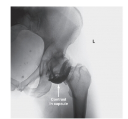
-Refer to the figure. Name this image.
A) Anteroposterior radiograph of the hip
B) Fluoroscopic image of the hip
C) CT of the hip
D) MRI of the hip
Question

-Refer to the figure. What is the diagnostic purpose of the contrast?
A) Evaluate the joint cartilage
B) Identify stress fracture
C) Determine vascularity
D) Stage a tumor
Question

-Refer to the figure. Name the image and view.
A) CT, sagittal
B) CT, coronal
C) MRI, sagittal
D) MRI, coronal
Question
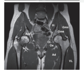
-Refer to the figure. What does the arrow point to?
A) Hip arthroplasty
B) Infection
C) Avascular necrosis
D) Fracture
Question
-You recognize the pathological condition in question above because of what characteristic?
A) Low signal line
B) High signal line
C) Metal hardware
D) Slipped femoral head
Question
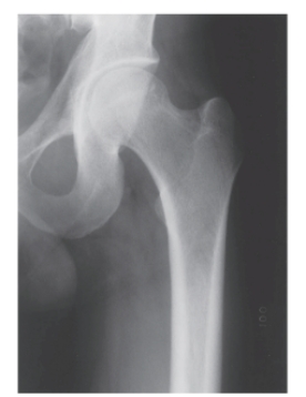
-Refer to the figure. Identify the radiographic view.
A) Anteroposterior radiograph of the hip
B) Lateral radiograph of the hip
C) Lateral "frog-leg" radiograph of the hip
D) Oblique view of the hip
Question

-Refer to the figure. What can be said about the image of cortical margins of the femur?
A) The thickness of the cortical image is normal
B) The absence of cortical image at the femoral neck indicates a demineralized state
C) The thickness of the cortical margins is pronounced, indicating a bone deposition disease
D) The pencil-thin cortical margins on the shaft indicate osteoporosis

Unlock Deck
Sign up to unlock the cards in this deck!
Unlock Deck
Unlock Deck
1/10
Play
Full screen (f)
Deck 12: Radiologic Evaluation of the Pelvis and Hip
1
Which of the following is true regarding the pelvis in males versus females?
A) Broader and greater angle of pubic arch
B) Broader and smaller angle of pubic arch
C) Narrower and greater angle of pubic arch
D) Narrower and smaller angle of pubic arch
A) Broader and greater angle of pubic arch
B) Broader and smaller angle of pubic arch
C) Narrower and greater angle of pubic arch
D) Narrower and smaller angle of pubic arch
Narrower and smaller angle of pubic arch
2
Axial migration of the femoral head is a finding associated with which of the following conditions as a result of the periarticular loss of bone density?
A) Acetabular labral tear
B) Ligamentum teres tear
C) Osteoarthritis
D) Rheumatoid arthritis
A) Acetabular labral tear
B) Ligamentum teres tear
C) Osteoarthritis
D) Rheumatoid arthritis
Rheumatoid arthritis
3
Which of the following pathological conditions is the most common adolescent hip disorder?
A) Developmental dysplasia of the hip
B) Egger's cysts
C) Legg-Calve-Perthes disease
D) Slipped capital femoral epiphysis
A) Developmental dysplasia of the hip
B) Egger's cysts
C) Legg-Calve-Perthes disease
D) Slipped capital femoral epiphysis
Slipped capital femoral epiphysis
4
-Refer to the figure. Name this image.
A) Anteroposterior radiograph of the hip
B) Fluoroscopic image of the hip
C) CT of the hip
D) MRI of the hip
Unlock Deck
Unlock for access to all 10 flashcards in this deck.
Unlock Deck
k this deck
5
-Refer to the figure. What is the diagnostic purpose of the contrast?
A) Evaluate the joint cartilage
B) Identify stress fracture
C) Determine vascularity
D) Stage a tumor
Unlock Deck
Unlock for access to all 10 flashcards in this deck.
Unlock Deck
k this deck
6
-Refer to the figure. Name the image and view.
A) CT, sagittal
B) CT, coronal
C) MRI, sagittal
D) MRI, coronal
Unlock Deck
Unlock for access to all 10 flashcards in this deck.
Unlock Deck
k this deck
7
-Refer to the figure. What does the arrow point to?
A) Hip arthroplasty
B) Infection
C) Avascular necrosis
D) Fracture
Unlock Deck
Unlock for access to all 10 flashcards in this deck.
Unlock Deck
k this deck
8
-You recognize the pathological condition in question above because of what characteristic?
A) Low signal line
B) High signal line
C) Metal hardware
D) Slipped femoral head
Unlock Deck
Unlock for access to all 10 flashcards in this deck.
Unlock Deck
k this deck
9
-Refer to the figure. Identify the radiographic view.
A) Anteroposterior radiograph of the hip
B) Lateral radiograph of the hip
C) Lateral "frog-leg" radiograph of the hip
D) Oblique view of the hip
Unlock Deck
Unlock for access to all 10 flashcards in this deck.
Unlock Deck
k this deck
10
-Refer to the figure. What can be said about the image of cortical margins of the femur?
A) The thickness of the cortical image is normal
B) The absence of cortical image at the femoral neck indicates a demineralized state
C) The thickness of the cortical margins is pronounced, indicating a bone deposition disease
D) The pencil-thin cortical margins on the shaft indicate osteoporosis
Unlock Deck
Unlock for access to all 10 flashcards in this deck.
Unlock Deck
k this deck
Unlock Deck
Unlock for access to all 10 flashcards in this deck.


