Deck 19: Image Evaluation
Full screen (f)
Question
Question
Question
Question
Question
Question
What is wrong with Figure 19-8?
A) There is too much density on the image.
B) The anatomy of interest is rotated.
C) Image contrast is too low.
D) An artifact is present on the image.
A) There is too much density on the image.
B) The anatomy of interest is rotated.
C) Image contrast is too low.
D) An artifact is present on the image.
Question
What projection and anatomy of interest are demonstrated in Figure 19-3?
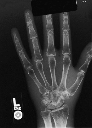
A) PA projection of the hand
B) AP projection of the thumb
C) Lateral projection of the hand
D) AP projection of the hand
A) PA projection of the hand
B) AP projection of the thumb
C) Lateral projection of the hand
D) AP projection of the hand
Question
Question
Question
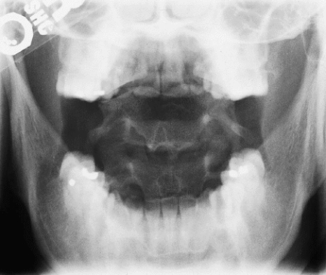
What projection and anatomy of interest are demonstrated in Figure 19-6?
A) AP projection of the forearm
B) AP projection of the elbow
C) Lateral projection of the forearm
D) Lateral projection of the elbow
A) AP projection of the forearm
B) AP projection of the elbow
C) Lateral projection of the forearm
D) Lateral projection of the elbow
Question
What error is evident in Figure 19-4?
A) Part of the anatomy of interest is not included on the image.
B) The anatomy of interest is rotated out of proper position.
C) There is no right or left side marker visible.
D) There is no error evident in image 19-4.
A) Part of the anatomy of interest is not included on the image.
B) The anatomy of interest is rotated out of proper position.
C) There is no right or left side marker visible.
D) There is no error evident in image 19-4.
Question
Question
Question
Question
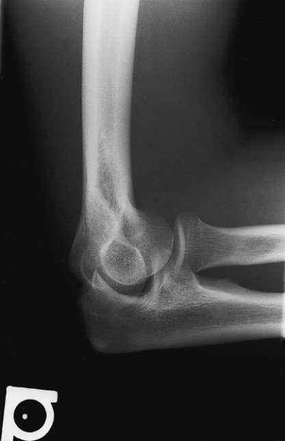
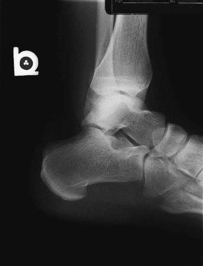
Which of the following errors is evident in Figure 19-5?
A) There is rotation of the anatomy of interest.
B) CR and IR were not aligned.
C) The image is too dark.
D) There is no error evident in Figure 19-5.
A) There is rotation of the anatomy of interest.
B) CR and IR were not aligned.
C) The image is too dark.
D) There is no error evident in Figure 19-5.
Question
What projection is demonstrated in Figure 19-2?
A) PA
B) PA oblique
C) AP
D) Lateral→
A) PA
B) PA oblique
C) AP
D) Lateral→
Question
Question
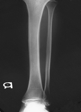
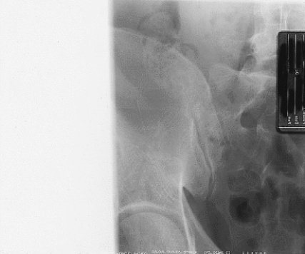
What is wrong with Figure 19-7?
A) Anatomy is rotated to the left.
B) A radiographic marker is not present.
C) There is too much density on the image.
D) The image contrast is too high.
A) Anatomy is rotated to the left.
B) A radiographic marker is not present.
C) There is too much density on the image.
D) The image contrast is too high.
Question
Question
What position is demonstrated in Figure 19-1?
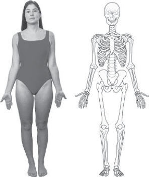
A) Recumbent
B) Anatomic
C) Decubitus
D) Prone
A) Recumbent
B) Anatomic
C) Decubitus
D) Prone

Unlock Deck
Sign up to unlock the cards in this deck!
Unlock Deck
Unlock Deck
1/20
Play
Full screen (f)
Deck 19: Image Evaluation
1
What error is evident in Figure 19-2?
A) The image is underexposed.
B) An artifact is present.
C) The radiation field is not centered over the anatomy.
D) Anatomy is not oriented correctly for this projection.
A) The image is underexposed.
B) An artifact is present.
C) The radiation field is not centered over the anatomy.
D) Anatomy is not oriented correctly for this projection.
The radiation field is not centered over the anatomy.
2
What is the first step in a systematic image review?
A) Check for accurate and complete image identification markers.
B) Check for pertinent and complete anatomy in the proper position.
C) Check for appropriate density and contrast.
D) Check for aesthetic quality.
A) Check for accurate and complete image identification markers.
B) Check for pertinent and complete anatomy in the proper position.
C) Check for appropriate density and contrast.
D) Check for aesthetic quality.
Check for accurate and complete image identification markers.
3
What is the proper method to hang an AP projection of the foot?
A) With the toes pointing toward the floor
B) With the toes pointing toward the ceiling
C) With the toes pointing toward the right
D) With the toes pointing toward the left
A) With the toes pointing toward the floor
B) With the toes pointing toward the ceiling
C) With the toes pointing toward the right
D) With the toes pointing toward the left
With the toes pointing toward the ceiling
4
Which of the following describes optimum radiographic viewing conditions?
A) Radiographs should be viewed with a "hot light."
B) Radiographs should be viewed in a totally darkened room.
C) Radiographs should be viewed in a normally lit room.
D) Radiographs should be viewed in a room with low light.
A) Radiographs should be viewed with a "hot light."
B) Radiographs should be viewed in a totally darkened room.
C) Radiographs should be viewed in a normally lit room.
D) Radiographs should be viewed in a room with low light.
Unlock Deck
Unlock for access to all 20 flashcards in this deck.
Unlock Deck
k this deck
5
Which of the following errors are evident in Figure 19-3?
1) The anatomy of interest is not centered to the IR.
2) There is too much density.
3) The blocker is in the anatomy of interest.
A) 1 and 2 only
B) 1 and 3 only
C) 2 and 3 only
D) 1, 2, and 3
1) The anatomy of interest is not centered to the IR.
2) There is too much density.
3) The blocker is in the anatomy of interest.
A) 1 and 2 only
B) 1 and 3 only
C) 2 and 3 only
D) 1, 2, and 3
Unlock Deck
Unlock for access to all 20 flashcards in this deck.
Unlock Deck
k this deck
6
What is wrong with Figure 19-8?
A) There is too much density on the image.
B) The anatomy of interest is rotated.
C) Image contrast is too low.
D) An artifact is present on the image.
A) There is too much density on the image.
B) The anatomy of interest is rotated.
C) Image contrast is too low.
D) An artifact is present on the image.
Unlock Deck
Unlock for access to all 20 flashcards in this deck.
Unlock Deck
k this deck
7
What projection and anatomy of interest are demonstrated in Figure 19-3?
A) PA projection of the hand
B) AP projection of the thumb
C) Lateral projection of the hand
D) AP projection of the hand
A) PA projection of the hand
B) AP projection of the thumb
C) Lateral projection of the hand
D) AP projection of the hand
Unlock Deck
Unlock for access to all 20 flashcards in this deck.
Unlock Deck
k this deck
8
What is the customary method to hang an image for viewing?
1) With the superior anatomy at the top
2) As if the patient were facing the viewer
3) As if the patient were lying down
A) 1 and 2 only
B) 1 and 3 only
C) 2 and 3 only
D) 1, 2, and 3
1) With the superior anatomy at the top
2) As if the patient were facing the viewer
3) As if the patient were lying down
A) 1 and 2 only
B) 1 and 3 only
C) 2 and 3 only
D) 1, 2, and 3
Unlock Deck
Unlock for access to all 20 flashcards in this deck.
Unlock Deck
k this deck
9
How can Figure 19-8 be corrected?
A) Decrease mAs by at least 50%.
B) Remove artifact from patient.
C) Decrease collimation.
D) Decrease kVp.
A) Decrease mAs by at least 50%.
B) Remove artifact from patient.
C) Decrease collimation.
D) Decrease kVp.
Unlock Deck
Unlock for access to all 20 flashcards in this deck.
Unlock Deck
k this deck
10
What projection and anatomy of interest are demonstrated in Figure 19-6?
A) AP projection of the forearm
B) AP projection of the elbow
C) Lateral projection of the forearm
D) Lateral projection of the elbow
A) AP projection of the forearm
B) AP projection of the elbow
C) Lateral projection of the forearm
D) Lateral projection of the elbow
Unlock Deck
Unlock for access to all 20 flashcards in this deck.
Unlock Deck
k this deck
11
What error is evident in Figure 19-4?
A) Part of the anatomy of interest is not included on the image.
B) The anatomy of interest is rotated out of proper position.
C) There is no right or left side marker visible.
D) There is no error evident in image 19-4.
A) Part of the anatomy of interest is not included on the image.
B) The anatomy of interest is rotated out of proper position.
C) There is no right or left side marker visible.
D) There is no error evident in image 19-4.
Unlock Deck
Unlock for access to all 20 flashcards in this deck.
Unlock Deck
k this deck
12
How can Figure 19-7 be corrected?
A) Reduce mAs by at least 50%.
B) Add radiographic marker.
C) Correct rotation of anatomy.
D) Reduce kVp by at least 50%.
A) Reduce mAs by at least 50%.
B) Add radiographic marker.
C) Correct rotation of anatomy.
D) Reduce kVp by at least 50%.
Unlock Deck
Unlock for access to all 20 flashcards in this deck.
Unlock Deck
k this deck
13
What causes an unexposed area at the top or bottom of a radiographic image?
A) Scatter radiation
B) Patient not aligned to the IR
C) CR not aligned to the IR
D) CR not centered to anatomy of interest
A) Scatter radiation
B) Patient not aligned to the IR
C) CR not aligned to the IR
D) CR not centered to anatomy of interest
Unlock Deck
Unlock for access to all 20 flashcards in this deck.
Unlock Deck
k this deck
14
Which of the following will improve the quality of Figure 19-3?
A) Center the IR and CR more distal and right on the patient.
B) Center the IR and CR more distal and left on the patient.
C) Center the IR and CR more proximal and right on the patient.
D) Center the IR and CR more proximal and left on the patient.
A) Center the IR and CR more distal and right on the patient.
B) Center the IR and CR more distal and left on the patient.
C) Center the IR and CR more proximal and right on the patient.
D) Center the IR and CR more proximal and left on the patient.
Unlock Deck
Unlock for access to all 20 flashcards in this deck.
Unlock Deck
k this deck
15
Which of the following errors is evident in Figure 19-5?
A) There is rotation of the anatomy of interest.
B) CR and IR were not aligned.
C) The image is too dark.
D) There is no error evident in Figure 19-5.
A) There is rotation of the anatomy of interest.
B) CR and IR were not aligned.
C) The image is too dark.
D) There is no error evident in Figure 19-5.
Unlock Deck
Unlock for access to all 20 flashcards in this deck.
Unlock Deck
k this deck
16
What projection is demonstrated in Figure 19-2?
A) PA
B) PA oblique
C) AP
D) Lateral→
A) PA
B) PA oblique
C) AP
D) Lateral→
Unlock Deck
Unlock for access to all 20 flashcards in this deck.
Unlock Deck
k this deck
17
What error is evident in Figure 19-6?
A)Artifacts are present.
B)Part of the anatomy of interest is not included on the image.
C)Anatomy of interest is not properly positioned.
D)The image contrast is too high.
A)Artifacts are present.
B)Part of the anatomy of interest is not included on the image.
C)Anatomy of interest is not properly positioned.
D)The image contrast is too high.
Unlock Deck
Unlock for access to all 20 flashcards in this deck.
Unlock Deck
k this deck
18
What is wrong with Figure 19-7?
A) Anatomy is rotated to the left.
B) A radiographic marker is not present.
C) There is too much density on the image.
D) The image contrast is too high.
A) Anatomy is rotated to the left.
B) A radiographic marker is not present.
C) There is too much density on the image.
D) The image contrast is too high.
Unlock Deck
Unlock for access to all 20 flashcards in this deck.
Unlock Deck
k this deck
19
Radiographs made with the patient in the ______ position are usually hung horizontally.
A) supine
B) prone
C) decubitus
D) upright
A) supine
B) prone
C) decubitus
D) upright
Unlock Deck
Unlock for access to all 20 flashcards in this deck.
Unlock Deck
k this deck
20
What position is demonstrated in Figure 19-1?
A) Recumbent
B) Anatomic
C) Decubitus
D) Prone
A) Recumbent
B) Anatomic
C) Decubitus
D) Prone
Unlock Deck
Unlock for access to all 20 flashcards in this deck.
Unlock Deck
k this deck
Unlock Deck
Unlock for access to all 20 flashcards in this deck.


