Deck 6: Lower Limb
Full screen (f)
Question
Question
Question
Question
Question
Question
Question
Question
Question
Question
Question
Which of these labeled structures or bones identifies the navicular? 
A)B
B)C
C)D
D)I
A)B
B)C
C)D
D)I
Question
Question
Question
Question
Question
Question
Question
Question
Question
Question
Question
This radiograph represents which of the following positions? 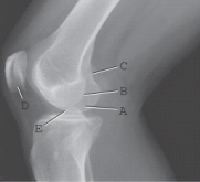
A)Mediolateral projection of the knee, but overextended
B)Mediolateral projection of the knee, but underrotated toward the IR
C)Mediolateral projection of the knee, but overrotated toward the IR
D)Correctly positioned mediolateral projection of the knee, but underexposed
A)Mediolateral projection of the knee, but overextended
B)Mediolateral projection of the knee, but underrotated toward the IR
C)Mediolateral projection of the knee, but overrotated toward the IR
D)Correctly positioned mediolateral projection of the knee, but underexposed
Question
Question
Question
Question
Question
Question
Which one of the labeled structures is the medial condyle of the femur? 
A)A
B)B
C)C
D)D
E)E
A)A
B)B
C)C
D)D
E)E
Question
Question
Which of these labeled structures or bones identifies the metatarsophalangeal joint? 
A)E
B)F
C)G
D)H
A)E
B)F
C)G
D)H
Question
Which of the labeled structures is the adductor tubercle? 
A)A
B)B
C)C
D)D
E)E
A)A
B)B
C)C
D)D
E)E
Question
Which of these labeled structures or bones identifies the talus? 
A)A
B)B
C)I
D)J
A)A
B)B
C)I
D)J
Question
Question
Question
Question
Which of these labeled structures or bones identifies the lateral (third) cuneiform? 
A)B
B)C
C)D
D)I
A)B
B)C
C)D
D)I
Question
Which projection and/or position of the foot is represented by this drawing of the foot? 
A)AP projection, no rotation
B)AP oblique, 15° to 20° medial rotation
C)AP oblique, 45° lateral rotation
D)AP oblique, 45° medial rotation
A)AP projection, no rotation
B)AP oblique, 15° to 20° medial rotation
C)AP oblique, 45° lateral rotation
D)AP oblique, 45° medial rotation
Question
Question
Question
Which of the labeled structures is the lateral condyle of the femur? 
A)A
B)B
C)C
D)D
E)E
A)A
B)B
C)C
D)D
E)E
Question
Question
Question
Question
Question
Question
Question
Question
Question
Question
Question
Question
Question
Question
Question
Question
Question
Question
Question
Question
Question
Question
Question
Question
Question
Question
Question
Question
Question
Question
Question
Question
Question
Question
Question
Question
Question
Question
Question
Question

Unlock Deck
Sign up to unlock the cards in this deck!
Unlock Deck
Unlock Deck
1/110
Play
Full screen (f)
Deck 6: Lower Limb
1
To decrease the angle between the anterior surface of the foot and anterior surface of the lower leg is described as:
A)plantar flexion.
B)inversion.
C)dorsiflexion.
D)eversion.
A)plantar flexion.
B)inversion.
C)dorsiflexion.
D)eversion.
dorsiflexion.
2
Which term describes the top or anterior surface of the foot?
A)Palmar
B)Dorsum
C)Volar
D)Plantar
A)Palmar
B)Dorsum
C)Volar
D)Plantar
Dorsum
3
Another term for the intercondylar sulcus is the:
A)articular facets.
B)patellar surface.
C)intercondylar fossa.
D)intercondylar recess.
A)articular facets.
B)patellar surface.
C)intercondylar fossa.
D)intercondylar recess.
patellar surface.
4
Which tendon attaches directly to the tibial tuberosity?
A)Patellar
B)Quadriceps
C)Soleus
D)Collateral
A)Patellar
B)Quadriceps
C)Soleus
D)Collateral
Unlock Deck
Unlock for access to all 110 flashcards in this deck.
Unlock Deck
k this deck
5
A tear of the tibial (medial) collateral ligament (MCL) caused by a trauma injury is frequently associated with tears of:
A)the anterior cruciate ligament (ACL) and the medial meniscus.
B)the posterior cruciate ligament (PCL) and the lateral meniscus.
C)the fibular lateral collateral ligament (LCL) and the patella ligament.
D)none of the above.
A)the anterior cruciate ligament (ACL) and the medial meniscus.
B)the posterior cruciate ligament (PCL) and the lateral meniscus.
C)the fibular lateral collateral ligament (LCL) and the patella ligament.
D)none of the above.
Unlock Deck
Unlock for access to all 110 flashcards in this deck.
Unlock Deck
k this deck
6
The medial malleolus is part of the:
A)talus.
B)calcaneus.
C)fibula.
D)tibia.
A)talus.
B)calcaneus.
C)fibula.
D)tibia.
Unlock Deck
Unlock for access to all 110 flashcards in this deck.
Unlock Deck
k this deck
7
What are the two arches of the foot?
A)Anterior and longitudinal
B)Longitudinal and transverse
C)Transverse and anterior
D)Instep and cross-step
A)Anterior and longitudinal
B)Longitudinal and transverse
C)Transverse and anterior
D)Instep and cross-step
Unlock Deck
Unlock for access to all 110 flashcards in this deck.
Unlock Deck
k this deck
8
The patellofemoral joint is a ____ joint with a ____ type of movement.
A)synovial; ginglymus
B)fibrous; immovable
C)synovial; saddle
D)synovial; bicondylar
A)synovial; ginglymus
B)fibrous; immovable
C)synovial; saddle
D)synovial; bicondylar
Unlock Deck
Unlock for access to all 110 flashcards in this deck.
Unlock Deck
k this deck
9
The ankle joint is a ____ joint with a ____ type of movement.
A)fibrous; plane
B)synovial; saddle
C)fibrous; ginglymus
D)synovial; ginglymus
A)fibrous; plane
B)synovial; saddle
C)fibrous; ginglymus
D)synovial; ginglymus
Unlock Deck
Unlock for access to all 110 flashcards in this deck.
Unlock Deck
k this deck
10
Saclike structures found in the knee joint that allow smooth articulation between ligaments and tendons are called:
A)bursae.
B)menisci.
C)synovial membranes.
D)synovial bodies.
A)bursae.
B)menisci.
C)synovial membranes.
D)synovial bodies.
Unlock Deck
Unlock for access to all 110 flashcards in this deck.
Unlock Deck
k this deck
11
Which of these labeled structures or bones identifies the navicular?
A)B
B)C
C)D
D)I
A)B
B)C
C)D
D)I
Unlock Deck
Unlock for access to all 110 flashcards in this deck.
Unlock Deck
k this deck
12
Which metatarsal bone of the foot has a prominent tuberosity most frequently fractured?
A)First
B)Third
C)Fourth
D)Fifth
A)First
B)Third
C)Fourth
D)Fifth
Unlock Deck
Unlock for access to all 110 flashcards in this deck.
Unlock Deck
k this deck
13
How many tarsal bones are found in the foot?
A)7
B)14
C)5
D)26
A)7
B)14
C)5
D)26
Unlock Deck
Unlock for access to all 110 flashcards in this deck.
Unlock Deck
k this deck
14
The best method of evaluating injuries to the menisci and ligaments of the knee joint involves:
A)stress views of the knee.
B)anteroposterior (AP), AP oblique, and lateral projections of the knee.
C)intercondylar fossa projections.
D)a magnetic resonance imaging procedure.
A)stress views of the knee.
B)anteroposterior (AP), AP oblique, and lateral projections of the knee.
C)intercondylar fossa projections.
D)a magnetic resonance imaging procedure.
Unlock Deck
Unlock for access to all 110 flashcards in this deck.
Unlock Deck
k this deck
15
How many articular facets make up the subtalar joint?
A)One
B)Two
C)Three
D)Four
A)One
B)Two
C)Three
D)Four
Unlock Deck
Unlock for access to all 110 flashcards in this deck.
Unlock Deck
k this deck
16
A radiographic appearance of a well-circumscribed lucency within bones describes:
A)gout.
B)Ewing's sarcoma.
C)a bone cyst.
D)Osgood-Schlatter disease.
A)gout.
B)Ewing's sarcoma.
C)a bone cyst.
D)Osgood-Schlatter disease.
Unlock Deck
Unlock for access to all 110 flashcards in this deck.
Unlock Deck
k this deck
17
The calcaneus articulates with the talus and the:
A)cuboid.
B)navicular.
C)medial cuneiform.
D)lateral cuneiform.
A)cuboid.
B)navicular.
C)medial cuneiform.
D)lateral cuneiform.
Unlock Deck
Unlock for access to all 110 flashcards in this deck.
Unlock Deck
k this deck
18
Which structure or bone contains the sustentaculum tali?
A)Calcaneus
B)Talus
C)Base of the fifth metatarsal
D)Tibia
A)Calcaneus
B)Talus
C)Base of the fifth metatarsal
D)Tibia
Unlock Deck
Unlock for access to all 110 flashcards in this deck.
Unlock Deck
k this deck
19
Where would the interphalangeal joint be found in the foot?
A)Between the phalanges of the second through fifth digits
B)Between the tarsal bones and phalanges
C)Between the phalanges of the first digit
D)Between any of the metatarsals and phalanges
A)Between the phalanges of the second through fifth digits
B)Between the tarsal bones and phalanges
C)Between the phalanges of the first digit
D)Between any of the metatarsals and phalanges
Unlock Deck
Unlock for access to all 110 flashcards in this deck.
Unlock Deck
k this deck
20
A radiographic appearance of a highly malignant and extensively destructive lesion that usually occurs in long bones and produces a sunburst pattern describes:
A)an osteomalacia.
B)an osteogenic sarcoma.
C)an osteoclastoma.
D)Reiter syndrome.
A)an osteomalacia.
B)an osteogenic sarcoma.
C)an osteoclastoma.
D)Reiter syndrome.
Unlock Deck
Unlock for access to all 110 flashcards in this deck.
Unlock Deck
k this deck
21
Which of the following joints is a modified ellipsoidal or condyloid joint?
A)Tarsometatarsal
B)Metatarsophalangeal
C)Proximal tibiofibular
D)Intertarsal
A)Tarsometatarsal
B)Metatarsophalangeal
C)Proximal tibiofibular
D)Intertarsal
Unlock Deck
Unlock for access to all 110 flashcards in this deck.
Unlock Deck
k this deck
22
This radiograph represents which of the following positions?
A)Mediolateral projection of the knee, but overextended
B)Mediolateral projection of the knee, but underrotated toward the IR
C)Mediolateral projection of the knee, but overrotated toward the IR
D)Correctly positioned mediolateral projection of the knee, but underexposed
A)Mediolateral projection of the knee, but overextended
B)Mediolateral projection of the knee, but underrotated toward the IR
C)Mediolateral projection of the knee, but overrotated toward the IR
D)Correctly positioned mediolateral projection of the knee, but underexposed
Unlock Deck
Unlock for access to all 110 flashcards in this deck.
Unlock Deck
k this deck
23
Which of the following routines should be performed for a study of the second toe?
A)AP, AP oblique with lateral rotation, mediolateral projection
B)AP, AP oblique with medial rotation, lateromedial projection
C)AP, AP oblique with lateral rotation, lateromedial projection
D)AP, AP oblique with medial rotation, mediolateral projection
A)AP, AP oblique with lateral rotation, mediolateral projection
B)AP, AP oblique with medial rotation, lateromedial projection
C)AP, AP oblique with lateral rotation, lateromedial projection
D)AP, AP oblique with medial rotation, mediolateral projection
Unlock Deck
Unlock for access to all 110 flashcards in this deck.
Unlock Deck
k this deck
24
To properly visualize the joint spaces with the AP projection of the foot, the CR must be:
A)parallel to the longitudinal arch.
B)perpendicular to the metatarsals.
C)perpendicular to the longitudinal arch.
D)parallel to the metatarsals.
A)parallel to the longitudinal arch.
B)perpendicular to the metatarsals.
C)perpendicular to the longitudinal arch.
D)parallel to the metatarsals.
Unlock Deck
Unlock for access to all 110 flashcards in this deck.
Unlock Deck
k this deck
25
What CR angulation is required for the AP medial oblique projection of the foot?
A)CR is perpendicular to the image receptor.
B)10° posterior
C)15° to 20° posterior
D)5° to 7° posterior
A)CR is perpendicular to the image receptor.
B)10° posterior
C)15° to 20° posterior
D)5° to 7° posterior
Unlock Deck
Unlock for access to all 110 flashcards in this deck.
Unlock Deck
k this deck
26
Extending the ankle joint or pointing the foot and toes downward is called:
A)dorsiflexion.
B)inversion.
C)eversion.
D)plantar flexion.
A)dorsiflexion.
B)inversion.
C)eversion.
D)plantar flexion.
Unlock Deck
Unlock for access to all 110 flashcards in this deck.
Unlock Deck
k this deck
27
How much is the foot dorsiflexed with the tangential projection for the sesamoid bones if the CR remains perpendicular to the image receptor?
A)15° to 20° from vertical
B)No flexion of the foot is required.
C)5° to 7° from vertical
D)30° to 45° from vertical
A)15° to 20° from vertical
B)No flexion of the foot is required.
C)5° to 7° from vertical
D)30° to 45° from vertical
Unlock Deck
Unlock for access to all 110 flashcards in this deck.
Unlock Deck
k this deck
28
Which one of the labeled structures is the medial condyle of the femur?
A)A
B)B
C)C
D)D
E)E
A)A
B)B
C)C
D)D
E)E
Unlock Deck
Unlock for access to all 110 flashcards in this deck.
Unlock Deck
k this deck
29
The distal tibiofibular joint is classified as a ____ joint.
A)synovial
B)fibrous
C)diarthrodial
D)synarthrodial
A)synovial
B)fibrous
C)diarthrodial
D)synarthrodial
Unlock Deck
Unlock for access to all 110 flashcards in this deck.
Unlock Deck
k this deck
30
Which of these labeled structures or bones identifies the metatarsophalangeal joint?
A)E
B)F
C)G
D)H
A)E
B)F
C)G
D)H
Unlock Deck
Unlock for access to all 110 flashcards in this deck.
Unlock Deck
k this deck
31
Which of the labeled structures is the adductor tubercle?
A)A
B)B
C)C
D)D
E)E
A)A
B)B
C)C
D)D
E)E
Unlock Deck
Unlock for access to all 110 flashcards in this deck.
Unlock Deck
k this deck
32
Which of these labeled structures or bones identifies the talus?
A)A
B)B
C)I
D)J
A)A
B)B
C)I
D)J
Unlock Deck
Unlock for access to all 110 flashcards in this deck.
Unlock Deck
k this deck
33
A mediolateral knee radiograph that is overrotated toward the image receptor can be recognized by which of the following?
A)The fibular head will appear more superimposed by the tibia than a true lateral.
B)The fibular head will appear less superimposed by the tibia than a true lateral.
C)The medial condyle of the femur will appear more posterior.
D)Both A and C are correct.
A)The fibular head will appear more superimposed by the tibia than a true lateral.
B)The fibular head will appear less superimposed by the tibia than a true lateral.
C)The medial condyle of the femur will appear more posterior.
D)Both A and C are correct.
Unlock Deck
Unlock for access to all 110 flashcards in this deck.
Unlock Deck
k this deck
34
How much central ray (CR) angulation (if any) should be used for an AP projection of the toes (without elevation of forefoot)?
A)Keep CR perpendicular to IR.
B)5° toward calcaneus
C)10° to 15° toward calcaneus
D)20° to 25° toward calcaneus
A)Keep CR perpendicular to IR.
B)5° toward calcaneus
C)10° to 15° toward calcaneus
D)20° to 25° toward calcaneus
Unlock Deck
Unlock for access to all 110 flashcards in this deck.
Unlock Deck
k this deck
35
Which position of the foot will best demonstrate the lateral (third) cuneiform?
A)AP oblique with medial rotation
B)AP oblique with lateral rotation
C)AP projection
D)Mediolateral projection
A)AP oblique with medial rotation
B)AP oblique with lateral rotation
C)AP projection
D)Mediolateral projection
Unlock Deck
Unlock for access to all 110 flashcards in this deck.
Unlock Deck
k this deck
36
Which of these labeled structures or bones identifies the lateral (third) cuneiform?
A)B
B)C
C)D
D)I
A)B
B)C
C)D
D)I
Unlock Deck
Unlock for access to all 110 flashcards in this deck.
Unlock Deck
k this deck
37
Which projection and/or position of the foot is represented by this drawing of the foot?
A)AP projection, no rotation
B)AP oblique, 15° to 20° medial rotation
C)AP oblique, 45° lateral rotation
D)AP oblique, 45° medial rotation
A)AP projection, no rotation
B)AP oblique, 15° to 20° medial rotation
C)AP oblique, 45° lateral rotation
D)AP oblique, 45° medial rotation
Unlock Deck
Unlock for access to all 110 flashcards in this deck.
Unlock Deck
k this deck
38
What is one advantage of the lateromedial projection of the foot?
A)It is more comfortable for the patient.
B)It better demonstrates the intertarsal joints.
C)The foot assumes a truer lateral position.
D)It opens the subtalar joint.
A)It is more comfortable for the patient.
B)It better demonstrates the intertarsal joints.
C)The foot assumes a truer lateral position.
D)It opens the subtalar joint.
Unlock Deck
Unlock for access to all 110 flashcards in this deck.
Unlock Deck
k this deck
39
How much CR angulation to the long axis of the foot is required for the plantodorsal (axial) projection of the calcaneus?
A)45° to 50°
B)15° to 20°
C)30° to 35°
D)40°
A)45° to 50°
B)15° to 20°
C)30° to 35°
D)40°
Unlock Deck
Unlock for access to all 110 flashcards in this deck.
Unlock Deck
k this deck
40
Which of the labeled structures is the lateral condyle of the femur?
A)A
B)B
C)C
D)D
E)E
A)A
B)B
C)C
D)D
E)E
Unlock Deck
Unlock for access to all 110 flashcards in this deck.
Unlock Deck
k this deck
41
A radiograph of an AP medial oblique projection of the foot, if positioned correctly, should demonstrate:
A)first through fifth metatarsals free of superimposition.
B)third through fifth metatarsals free of superimposition.
C)first and second cuneiform joint space is open.
D)CR is centered to midshaft of third metatarsal.
A)first through fifth metatarsals free of superimposition.
B)third through fifth metatarsals free of superimposition.
C)first and second cuneiform joint space is open.
D)CR is centered to midshaft of third metatarsal.
Unlock Deck
Unlock for access to all 110 flashcards in this deck.
Unlock Deck
k this deck
42
A radiograph of an AP ankle projection reveals that the lateral joint space is not open (lateral malleolus is partially superimposed by the talus).The superior and medial joint spaces are open.What should the technologist do to correct this problem and improve the image?
A)Rotate the ankle more laterally.
B)Rotate the ankle more medially.
C)Nothing; this is an acceptable image.
D)Dorsiflex the foot.
A)Rotate the ankle more laterally.
B)Rotate the ankle more medially.
C)Nothing; this is an acceptable image.
D)Dorsiflex the foot.
Unlock Deck
Unlock for access to all 110 flashcards in this deck.
Unlock Deck
k this deck
43
A radiograph of an AP projection of the second toe reveals that the interphalangeal joints are not open.What is the most likely cause for this radiographic outcome?
A)Rotation of the toes.
B)Excessive SID was used.
C)AP projection was made; should have performed the PA projection.
D)Incorrect or inadequate CR centering or angle.
A)Rotation of the toes.
B)Excessive SID was used.
C)AP projection was made; should have performed the PA projection.
D)Incorrect or inadequate CR centering or angle.
Unlock Deck
Unlock for access to all 110 flashcards in this deck.
Unlock Deck
k this deck
44
Which of the following projections of the ankle will best demonstrate the open joint space of the lateral aspect of the ankle joint?
A)AP oblique with 45° rotation
B)AP mortise projection
C)Lateromedial ankle
D)AP projection
A)AP oblique with 45° rotation
B)AP mortise projection
C)Lateromedial ankle
D)AP projection
Unlock Deck
Unlock for access to all 110 flashcards in this deck.
Unlock Deck
k this deck
45
What CR angle should be used for a mediolateral projection of the knee on a short, wide-pelvis patient?
A)No CR angle is required.
B)5° cephalad
C)7° to 10° cephalad
D)5° to 10° caudad
A)No CR angle is required.
B)5° cephalad
C)7° to 10° cephalad
D)5° to 10° caudad
Unlock Deck
Unlock for access to all 110 flashcards in this deck.
Unlock Deck
k this deck
46
Which special position of the knee requires that the patient be placed supine with 40° flexion of the knee and the CR angled 30° from the long axis of the femur?
A)Bilateral Merchant method
B)PA axial, Camp-Coventry method
C)PA axial, Holmblad method
D)Tangential, Hughston method
A)Bilateral Merchant method
B)PA axial, Camp-Coventry method
C)PA axial, Holmblad method
D)Tangential, Hughston method
Unlock Deck
Unlock for access to all 110 flashcards in this deck.
Unlock Deck
k this deck
47
How much flexion of the knee is recommended for the lateral projection of the patella?
A)5° to 10°
B)20° to 30°
C)35° to 40°
D)45° to 50°
A)5° to 10°
B)20° to 30°
C)35° to 40°
D)45° to 50°
Unlock Deck
Unlock for access to all 110 flashcards in this deck.
Unlock Deck
k this deck
48
What is the major disadvantage of using 45° of flexion for the mediolateral projection of the knee?
A)Draws the patella into the intercondylar sulcus.
B)May cause injury to the anterior cruciate ligament.
C)Prevents superimposition of the distal aspect of the femoral condyles.
D)Can distort any visible fat pads.
A)Draws the patella into the intercondylar sulcus.
B)May cause injury to the anterior cruciate ligament.
C)Prevents superimposition of the distal aspect of the femoral condyles.
D)Can distort any visible fat pads.
Unlock Deck
Unlock for access to all 110 flashcards in this deck.
Unlock Deck
k this deck
49
Which projection of the knee will best demonstrate the neck of the fibula without superimposition of the tibia?
A)AP
B)Lateral
C)AP oblique with medial rotation
D)AP oblique with lateral rotation
A)AP
B)Lateral
C)AP oblique with medial rotation
D)AP oblique with lateral rotation
Unlock Deck
Unlock for access to all 110 flashcards in this deck.
Unlock Deck
k this deck
50
Where is the CR placed for a mediolateral projection of the calcaneus?
A)Base of the fifth metatarsal
B)Trochlear process
C)Base of the third metatarsal
D)1 inch (2.5 cm) inferior to medial malleolus
A)Base of the fifth metatarsal
B)Trochlear process
C)Base of the third metatarsal
D)1 inch (2.5 cm) inferior to medial malleolus
Unlock Deck
Unlock for access to all 110 flashcards in this deck.
Unlock Deck
k this deck
51
What CR angulation is recommended for an AP projection of the knee on a patient with an ASIS-to-tabletop measurement of 18 cm?
A)3° to 5° caudad
B)CR is perpendicular to the IR.
C)3° to 5° cephalad
D)10° to 15° cephalad
A)3° to 5° caudad
B)CR is perpendicular to the IR.
C)3° to 5° cephalad
D)10° to 15° cephalad
Unlock Deck
Unlock for access to all 110 flashcards in this deck.
Unlock Deck
k this deck
52
The profile appearance of the adductor tubercle and excessive superimposition of the fibular head and neck on a mediolateral knee projection indicate:
A)overrotation of the knee toward the IR.
B)underrotation of the knee toward the IR.
C)a true lateral knee.
D)the CR should be angled 5° to 7° cephalad.
A)overrotation of the knee toward the IR.
B)underrotation of the knee toward the IR.
C)a true lateral knee.
D)the CR should be angled 5° to 7° cephalad.
Unlock Deck
Unlock for access to all 110 flashcards in this deck.
Unlock Deck
k this deck
53
The purpose of the AP stress views of the ankle is to demonstrate:
A)possible stress fractures.
B)possible joint separations or ligament tears.
C)loose bodies in ankle joints.
D)tears in the joint meniscus.
A)possible stress fractures.
B)possible joint separations or ligament tears.
C)loose bodies in ankle joints.
D)tears in the joint meniscus.
Unlock Deck
Unlock for access to all 110 flashcards in this deck.
Unlock Deck
k this deck
54
A radiograph of a mediolateral projection of the patella reveals that the femoropatellar joint space is not open.The patella is within the intercondylar sulcus.The most likely cause of this is:
A)excessive extension of the knee.
B)excessive angulation of the CR.
C)insufficient angulation of the CR.
D)excessive flexion of the knee.
A)excessive extension of the knee.
B)excessive angulation of the CR.
C)insufficient angulation of the CR.
D)excessive flexion of the knee.
Unlock Deck
Unlock for access to all 110 flashcards in this deck.
Unlock Deck
k this deck
55
How much rotation from an AP position of the ankle will typically produce an AP mortise projection?
A)No rotation is necessary.
B)45° to 60° lateral
C)15° to 20° medial
D)25° to 30° medial
A)No rotation is necessary.
B)45° to 60° lateral
C)15° to 20° medial
D)25° to 30° medial
Unlock Deck
Unlock for access to all 110 flashcards in this deck.
Unlock Deck
k this deck
56
What is the recommended SID for the superoinferior sitting tangential (Hobbs modification) method?
A)30 inches (77 cm)
B)40 inches (102 cm)
C)48 to 50 inches (123 to 128 cm)
D)72 inches (183 cm)
A)30 inches (77 cm)
B)40 inches (102 cm)
C)48 to 50 inches (123 to 128 cm)
D)72 inches (183 cm)
Unlock Deck
Unlock for access to all 110 flashcards in this deck.
Unlock Deck
k this deck
57
A radiograph of an AP knee reveals rotation with almost total superimposition of the fibular head and the proximal tibia.What must the technologist do to correct this positioning error on the repeat exposure?
A)Rotate the knee laterally slightly.
B)Rotate the knee medially slightly.
C)Angle the CR slightly more cephalad.
D)Nothing; this is an acceptable image.
A)Rotate the knee laterally slightly.
B)Rotate the knee medially slightly.
C)Angle the CR slightly more cephalad.
D)Nothing; this is an acceptable image.
Unlock Deck
Unlock for access to all 110 flashcards in this deck.
Unlock Deck
k this deck
58
What type of CR angle is required for the PA axial weight-bearing bilateral knee projection (Rosenberg method)?
A)10° caudad
B)5° to 7° cephalad
C)20° to 25° caudad
D)None.CR is perpendicular to IR.
A)10° caudad
B)5° to 7° cephalad
C)20° to 25° caudad
D)None.CR is perpendicular to IR.
Unlock Deck
Unlock for access to all 110 flashcards in this deck.
Unlock Deck
k this deck
59
Which joint surfaces of the ankle joint are most commonly open with an AP projection of the ankle?
A)Medial and superior
B)Lateral and medial
C)Superior and lateral
D)Medial, superior, and lateral
A)Medial and superior
B)Lateral and medial
C)Superior and lateral
D)Medial, superior, and lateral
Unlock Deck
Unlock for access to all 110 flashcards in this deck.
Unlock Deck
k this deck
60
To ensure that both joints are included on an AP projection of the tibia and fibula on an adult, the technologist can:
A)increase the SID to 60 inches (150 cm).
B)use a Bucky tray.
C)turn the image receptor diagonally to the lower leg.
D)use a tabletop technique.
A)increase the SID to 60 inches (150 cm).
B)use a Bucky tray.
C)turn the image receptor diagonally to the lower leg.
D)use a tabletop technique.
Unlock Deck
Unlock for access to all 110 flashcards in this deck.
Unlock Deck
k this deck
61
A radiograph of a plantodorsal (axial) projection of the calcaneus reveals foreshortening.The technologist used 60 kV, 6 mAs, 40-inch (102-cm) SID, and a 25° cephalad CR angle from the long axis of the foot.Which of the following modifications will produce a more diagnostic image of the calcaneus?
A)Plantarflex the foot.
B)Increase CR angulation.
C)Decrease CR angulation.
D)Increase kV to 70.
A)Plantarflex the foot.
B)Increase CR angulation.
C)Decrease CR angulation.
D)Increase kV to 70.
Unlock Deck
Unlock for access to all 110 flashcards in this deck.
Unlock Deck
k this deck
62
Which one of the following projections will best demonstrate signs of Osgood-Schlatter disease?
A)Plantodorsal (axial) and lateral calcaneus
B)AP, lateral, and oblique ankle
C)AP bilateral weight-bearing knees
D)AP and lateral knee
A)Plantodorsal (axial) and lateral calcaneus
B)AP, lateral, and oblique ankle
C)AP bilateral weight-bearing knees
D)AP and lateral knee
Unlock Deck
Unlock for access to all 110 flashcards in this deck.
Unlock Deck
k this deck
63
A patient comes to radiology with a clinical history of osteoarthritis of both knees.The referring physician wants a projection to evaluate the damage to the articular facets.Which of the following projections will provide the best image of this region of the knee?
A)Tangential projection (Hughston method)
B)AP axial projection (Béclere method)
C)PA axial weight-bearing bilateral knee projection (Rosenberg method)
D)Tangential projection (Settegast method)
A)Tangential projection (Hughston method)
B)AP axial projection (Béclere method)
C)PA axial weight-bearing bilateral knee projection (Rosenberg method)
D)Tangential projection (Settegast method)
Unlock Deck
Unlock for access to all 110 flashcards in this deck.
Unlock Deck
k this deck
64
A radiograph of an AP oblique foot with medial rotation demonstrates considerable superimposition of the third through fifth metatarsals.How must the original position be changed to eliminate this problem?
A)Increase obliquity of the foot.
B)Decrease obliquity of the foot.
C)Increase CR angle.
D)Decrease CR angle.
A)Increase obliquity of the foot.
B)Decrease obliquity of the foot.
C)Increase CR angle.
D)Decrease CR angle.
Unlock Deck
Unlock for access to all 110 flashcards in this deck.
Unlock Deck
k this deck
65
Which of the following imaging modalities and/or procedures will provide the best assessment for osteomyelitis of the foot?
A)Nuclear medicine
B)Ultrasound
C)Computed tomography
D)Arthrography
A)Nuclear medicine
B)Ultrasound
C)Computed tomography
D)Arthrography
Unlock Deck
Unlock for access to all 110 flashcards in this deck.
Unlock Deck
k this deck
66
A patient comes to the radiology department for a knee study with special interest in the region of the proximal tibiofibular joint and the lateral condyle of the tibia.Which of the following positioning routines should the technologist obtain?
A)AP and lateral knee
B)AP, lateral, and lateral oblique knee
C)AP, lateral, and medial oblique knee
D)AP, lateral, and PA axial intercondylar fossa
A)AP and lateral knee
B)AP, lateral, and lateral oblique knee
C)AP, lateral, and medial oblique knee
D)AP, lateral, and PA axial intercondylar fossa
Unlock Deck
Unlock for access to all 110 flashcards in this deck.
Unlock Deck
k this deck
67
For the AP weight-bearing knee projection on an average patient, the CR should be:
A)10° caudad.
B)5° to 10° cephalad.
C)perpendicular to the image receptor.
D)perpendicular to the image receptor, but SID should be increased to 60 inches (153 cm).
A)10° caudad.
B)5° to 10° cephalad.
C)perpendicular to the image receptor.
D)perpendicular to the image receptor, but SID should be increased to 60 inches (153 cm).
Unlock Deck
Unlock for access to all 110 flashcards in this deck.
Unlock Deck
k this deck
68
A patient comes to radiology with an infection involving the sesamoid bones of the foot.Beyond the routine foot projections, which one of the following projections can be performed to best demonstrate these structures?
A)PA axial Camp-Coventry method
B)AP weight-bearing foot projection
C)Lateral weight-bearing projection
D)Tangential projection
A)PA axial Camp-Coventry method
B)AP weight-bearing foot projection
C)Lateral weight-bearing projection
D)Tangential projection
Unlock Deck
Unlock for access to all 110 flashcards in this deck.
Unlock Deck
k this deck
69
A radiograph of an AP mortise projection of the ankle reveals that the lateral malleolus is slightly superimposed over the talus and the lateral joint space is not open.What is most likely cause for this radiographic outcome?
A)Excessive medial rotation of the foot and ankle
B)Insufficient medial rotation of the foot and ankle
C)Excessive plantar flexion of the foot and ankle
D)Excessive dorsiflexion of the foot and ankle
A)Excessive medial rotation of the foot and ankle
B)Insufficient medial rotation of the foot and ankle
C)Excessive plantar flexion of the foot and ankle
D)Excessive dorsiflexion of the foot and ankle
Unlock Deck
Unlock for access to all 110 flashcards in this deck.
Unlock Deck
k this deck
70
A patient enters the ED with an injury near the base of the first and second metatarsals.The basic foot projections are inconclusive on demonstrating a fracture to the medial cuneiform.Which of the following projections would best demonstrate this bone?
A)AP oblique with increased medial rotation
B)AP oblique with lateral rotation
C)AP weight-bearing projection
D)Lateral weight-bearing projection
A)AP oblique with increased medial rotation
B)AP oblique with lateral rotation
C)AP weight-bearing projection
D)Lateral weight-bearing projection
Unlock Deck
Unlock for access to all 110 flashcards in this deck.
Unlock Deck
k this deck
71
A patient comes to radiology with a history of chondromalacia of the patella.The orthopedic surgeon is concerned about possible loose bodies within the patellofemoral joint space.She wants the best projection to demonstrate this joint space.What projection should be performed?
A)Camp-Coventry method
B)Settegast method
C)AP axial projection
D)Merchant method
A)Camp-Coventry method
B)Settegast method
C)AP axial projection
D)Merchant method
Unlock Deck
Unlock for access to all 110 flashcards in this deck.
Unlock Deck
k this deck
72
The radiographic hallmark of Reiter's syndrome seen in young men is:
A)asymmetrical narrowing of femoropatellar joint.
B)destruction of the patella.
C)fluid in the joints of the foot.
D)erosion of the Achilles tendon insertion.
A)asymmetrical narrowing of femoropatellar joint.
B)destruction of the patella.
C)fluid in the joints of the foot.
D)erosion of the Achilles tendon insertion.
Unlock Deck
Unlock for access to all 110 flashcards in this deck.
Unlock Deck
k this deck
73
A patient comes to radiology with a history of chondromalacia of the patella.Her physician orders a projection of the patellofemoral joint space.Due to advanced emphysema, the patient cannot lie recumbent for this projection.Which of the following projections would be best for this patient?
A)Tangential projection-Settegast method
B)Tangential projection-Merchant method
C)AP axial projection-Béclere method
D)Superoinferior sitting tangential method-Hobbs modification
A)Tangential projection-Settegast method
B)Tangential projection-Merchant method
C)AP axial projection-Béclere method
D)Superoinferior sitting tangential method-Hobbs modification
Unlock Deck
Unlock for access to all 110 flashcards in this deck.
Unlock Deck
k this deck
74
A patient comes to radiology with a clinical history of a Lisfranc joint injury.Which of the following projections would best demonstrate this condition?
A)Weight-bearing foot series
B)AP weight-bearing knee projection
C)Tangential projection for patella
D)AP stress views of the ankle
A)Weight-bearing foot series
B)AP weight-bearing knee projection
C)Tangential projection for patella
D)AP stress views of the ankle
Unlock Deck
Unlock for access to all 110 flashcards in this deck.
Unlock Deck
k this deck
75
A patient enters radiology with a possible ligament tear to the lateral aspect of the ankle.Initial ankle radiographs are negative for fracture or dislocation.Because the clinic is in a rural setting, the patient cannot have an MRI performed to evaluate the ligaments of the ankle.Which of the following techniques may provide an assessment of the soft tissue structures of the ankle?
A)AP weight-bearing projections
B)AP mortise projection
C)AP stress positions
D)Axial plantodorsal projection
A)AP weight-bearing projections
B)AP mortise projection
C)AP stress positions
D)Axial plantodorsal projection
Unlock Deck
Unlock for access to all 110 flashcards in this deck.
Unlock Deck
k this deck
76
A patient enters the emergency department (ED) with a possible transverse fracture of the patella.Which of the following routines would safely provide the best images of the patella?
A)AP and horizontal beam lateral, no flexion of knee
B)AP and 5° to 10° flexion lateral
C)AP and Merchant method (tangential projection)
D)PA and 45° PA oblique with medial rotation
A)AP and horizontal beam lateral, no flexion of knee
B)AP and 5° to 10° flexion lateral
C)AP and Merchant method (tangential projection)
D)PA and 45° PA oblique with medial rotation
Unlock Deck
Unlock for access to all 110 flashcards in this deck.
Unlock Deck
k this deck
77
For the AP weight-bearing feet projection, the CR should be:
A)perpendicular to the image receptor.
B)angled 15° posteriorly.
C)directed horizontal.
D)angled 5° posteriorly.
A)perpendicular to the image receptor.
B)angled 15° posteriorly.
C)directed horizontal.
D)angled 5° posteriorly.
Unlock Deck
Unlock for access to all 110 flashcards in this deck.
Unlock Deck
k this deck
78
A geriatric patient comes to the radiology department for a study of the knee.The patient is unsteady and unsure of himself.Which intercondylar fossa projection would provide the best results without risk of injury to the patient?
A)Holmblad method
B)Hughston method
C)Camp-Coventry method
D)Rosenberg method
A)Holmblad method
B)Hughston method
C)Camp-Coventry method
D)Rosenberg method
Unlock Deck
Unlock for access to all 110 flashcards in this deck.
Unlock Deck
k this deck
79
A patient comes to radiology for an evaluation of the longitudinal arch of the foot.Which of the following projections would provide the best information about the arch?
A)Routine foot series
B)Plantodorsal (axial) projection
C)AP and lateral weight-bearing projections of foot
D)Sesamoid bone series projection
A)Routine foot series
B)Plantodorsal (axial) projection
C)AP and lateral weight-bearing projections of foot
D)Sesamoid bone series projection
Unlock Deck
Unlock for access to all 110 flashcards in this deck.
Unlock Deck
k this deck
80
A radiograph of a PA axial projection for the intercondylar fossa (Camp-Coventry method) does not demonstrate the fossa well.It is foreshortened.The following positioning factors were used: patient prone, knee flexed 40° to 45°, CR angled to be perpendicular to the femur, 40-inch SID, and no rotation of the lower limb.Based on the factors used, what changes need to be made to produce a more diagnostic image?
A)Increase SID to at least 48 inches (123 cm).
B)CR must be perpendicular to lower leg.
C)Rotate lower extremity 10° medially.
D)Reduce flexion of the knee to 20° to 30°.
A)Increase SID to at least 48 inches (123 cm).
B)CR must be perpendicular to lower leg.
C)Rotate lower extremity 10° medially.
D)Reduce flexion of the knee to 20° to 30°.
Unlock Deck
Unlock for access to all 110 flashcards in this deck.
Unlock Deck
k this deck
Unlock Deck
Unlock for access to all 110 flashcards in this deck.


