Deck 20: The Cardiovascular System: The Heart
Full screen (f)
Question
Question
Question
Question
Question
Question
Question
Question
Question
Question
Question
Question
Question
Question
Question
Question
Question
Question
Question
Question
Question
Question
Question
Question
Question
Question
Question
Question
Question
Question
Question
Question
Question
Question
Question
This portion of the heart wall is responsible for the pumping action. 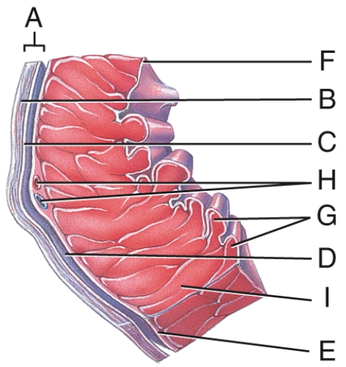
A)E
B)F
C)G
D)H
E)I
A)E
B)F
C)G
D)H
E)I
Question
Question
Question
Question
Question
Question
This is comprised of a thin layer of endothelium overlying a thin layer of connective tissue. 
A)C
B)D
C)E
D)F
E)G
A)C
B)D
C)E
D)F
E)G
Question
In the diagram,which labeled structure is the marginal branch of the right coronary artery? 
A)A
B)B
C)D
D)E
E)F
A)A
B)B
C)D
D)E
E)F
Question
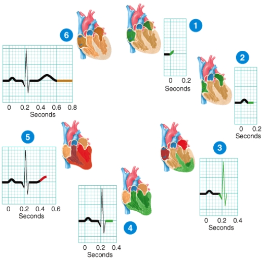
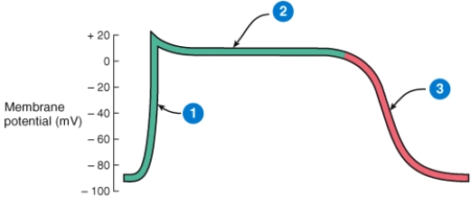
Which phases of a heartbeat shown in the diagram involve repolarization of the heart's four chambers?

a)1 and 4
b)2 and 4
c)4 and 6
d)1,3,and 5
e)1,2,4 and 6
f)3 and 5
a)1 and 4
b)2 and 4
c)4 and 6
d)1,3,and 5
e)1,2,4 and 6
f)3 and 5
Question
In the diagram,which labeled structure is the circumflex branch of the left coronary artery? 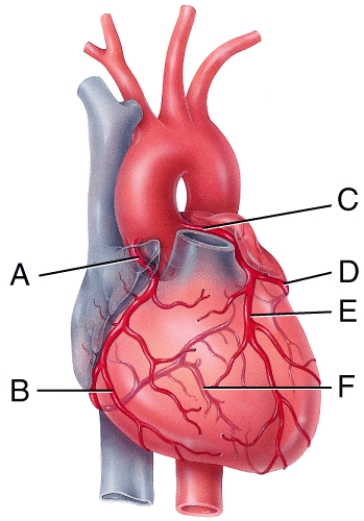
A)B
B)D
C)E
D)F
E)C
A)B
B)D
C)E
D)F
E)C
Question
In the diagram,which labeled structure is the pulmonary semilunar valve? 
A)B
B)D
C)E
D)A
E)None of these choices
A)B
B)D
C)E
D)A
E)None of these choices
Question
In the diagram,which labeled structure is the anterior interventricular branch of the left coronary artery? 
A)B
B)C
C)D
D)E
E)F
A)B
B)C
C)D
D)E
E)F
Question
In the diagram,where is the ascending aorta? 
A)A
B)B
C)D
D)F
E)H
A)A
B)B
C)D
D)F
E)H
Question
Which layer of the pericardium consists of dense irregular connective tissue? 
A)F
B)B
C)C
D)D
E)E
A)F
B)B
C)C
D)D
E)E
Question
Which of the labeled diagrams shows an atrial septal defect? 
A)A
B)B
C)C
D)D
E)E
A)A
B)B
C)C
D)D
E)E
Question
In the diagram,which labeled structure prevents blood flow from the right ventricle back into the right atrium? 
A)A
B)B
C)C
D)D
E)E
A)A
B)B
C)C
D)D
E)E
Question
In the diagram,which labeled structures are atrioventricular valves? 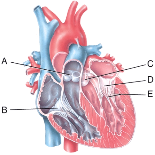
A)B only
B)D only
C)A and C
D)B and D
E)A,B,C and D
A)B only
B)D only
C)A and C
D)B and D
E)A,B,C and D
Question
Briefly describe what is happening at stage of the ECG labeled 5 in the diagram.
Question
In the diagram,where is the coronary sulcus? 
A)C
B)E
C)G
D)H
E)I
A)C
B)E
C)G
D)H
E)I
Question
In the diagram,where is the left auricle of the left atrium? 
A)C
B)F
C)G
D)H
E)I
A)C
B)F
C)G
D)H
E)I
Question
Which of the labeled steps in the diagram represent formation of the endocardial tubes? 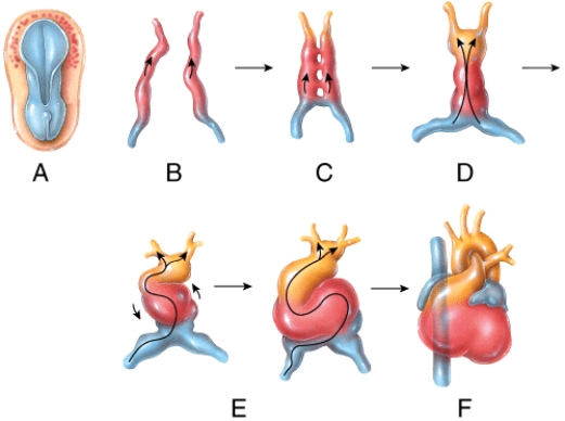
A)A
B)B
C)C
D)D
E)E
A)A
B)B
C)C
D)D
E)E
Question
In the diagram,these structures contain coronary blood vessels and a variable amount of fat. 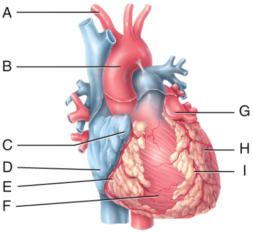
A)F and H
B)A and B
C)C and G
D)E and I
E)D and F
A)F and H
B)A and B
C)C and G
D)E and I
E)D and F
Question
Which of the labeled steps in the diagram represent formation of the primitive heart tube? 
A)A
B)B
C)C
D)D
E)E
A)A
B)B
C)C
D)D
E)E
Question
Describe what is happening during the phase of the cardiac action potential labeled 2 in the diagram.
Question
Which of the labeled diagrams shows coarctation of the aorta? 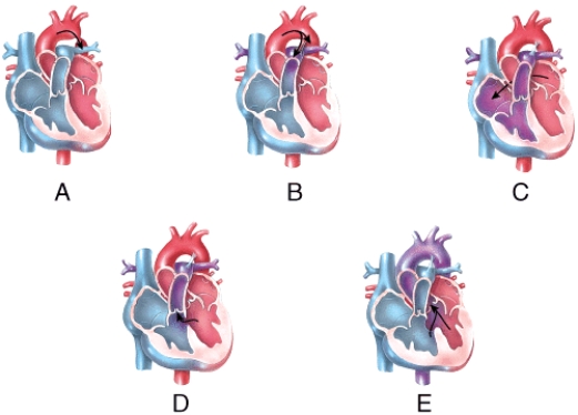
A)A
B)B
C)C
D)D
E)E
A)A
B)B
C)C
D)D
E)E
Question
In the diagram,where are the trabeculae carneae? 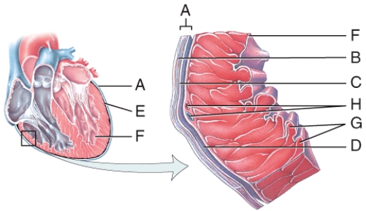
A)D
B)E
C)F
D)G
E)H
A)D
B)E
C)F
D)G
E)H
Question
What labeled structure in the figure increases the blood volume capacity of an atrium? 
A)B
B)G
C)D
D)F
A)B
B)G
C)D
D)F
Question
Which labeled structure in the figure carries the cardiac action potential directly into the contractile fibers of the ventricular myocardium? 
A)A
B)B
C)C
D)D
E)E
A)A
B)B
C)C
D)D
E)E
Question
Which labeled blood vessel in the diagram is an artery carrying deoxygenated blood? 
A)A
B)B
C)C
D)E
E)I
A)A
B)B
C)C
D)E
E)I
Question
Which blood vessel shown in the diagram is the left subclavian artery? 
A)A
B)B
C)E
D)F
E)H
A)A
B)B
C)E
D)F
E)H
Question
Which labeled blood vessel shown in the diagram is the left common carotid artery? 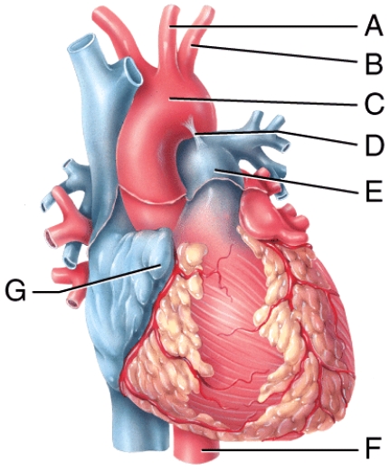
A)A
B)B
C)E
D)F
E)H
A)A
B)B
C)E
D)F
E)H
Question
Which blood vessel shown in the figure carries oxygenated blood to the lower thoracic cavity and the abdominal cavity of the body? 
A)A
B)B
C)E
D)F
E)H
A)A
B)B
C)E
D)F
E)H
Question
Which labeled blood vessel shown in the diagram is the right common carotid artery? 
A)A
B)B
C)E
D)F
E)not shown in the diagram
A)A
B)B
C)E
D)F
E)not shown in the diagram
Question
What labeled structure in the figure is the ligamentum arteriosum? 
A)A
B)B
C)C
D)D
A)A
B)B
C)C
D)D
Question
Which structure in the figure is labeled B? 
A)left common carotid artery
B)left subclavian artery
C)left pulmonary vein
D)mitral valve
A)left common carotid artery
B)left subclavian artery
C)left pulmonary vein
D)mitral valve
Question
Which labeled structure in the figure acts as the natural pacemaker of the heart? 
A)A
B)B
C)C
D)D
E)E
A)A
B)B
C)C
D)D
E)E
Question
Which labeled blood vessel carries oxygenated blood from the right lung back to the heart? 
A)B
B)K
C)J
D)E
E)C
A)B
B)K
C)J
D)E
E)C
Question
Which labeled structure shown in the diagram is a remnant of fetal circulation that is not directly involved in adult circulation? 
A)A
B)B
C)H
D)D
E)E
A)A
B)B
C)H
D)D
E)E
Question
Which labeled structure in the figure represents the only potential pathway for conducting action potentials from the atria to the ventricles? 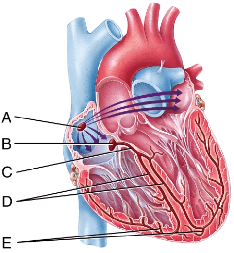
A)A
B)B
C)C
D)D
E)E
A)A
B)B
C)C
D)D
E)E
Question
Which labeled structure in the figure receives deoxygenated blood from the blood vessel labeled A? 
A)G
B)C
C)D
D)I
E)F
A)G
B)C
C)D
D)I
E)F
Question
What labeled structure in the figure is the descending aorta? 
A)F
B)C
C)D
D)A
A)F
B)C
C)D
D)A
Question
Which structure in the figure is labeled C? 
A)arch of aorta
B)pulmonary trunk
C)tricuspid valve
D)aortic valve
A)arch of aorta
B)pulmonary trunk
C)tricuspid valve
D)aortic valve
Question
Which structure in the figure is labeled A? 
A)left common carotid artery
B)left subclavian artery
C)left pulmonary vein
D)mitral valve
A)left common carotid artery
B)left subclavian artery
C)left pulmonary vein
D)mitral valve
Question
What labeled structure in the figure divides into the right and left pulmonary arteries to carry blood to the lungs? 
A)E
B)A
C)D
D)G
A)E
B)A
C)D
D)G
Question
Which labeled structure in the figure is the AV node? 
A)A
B)B
C)C
D)D
E)E
A)A
B)B
C)C
D)D
E)E
Question
Which labeled structure shown in the diagram is a pouch-like extension that serves to slightly increase the capacity of an atrium? 
A)F
B)E
C)G
D)I
E)D
A)F
B)E
C)G
D)I
E)D

Unlock Deck
Sign up to unlock the cards in this deck!
Unlock Deck
Unlock Deck
1/93
Play
Full screen (f)
Deck 20: The Cardiovascular System: The Heart
1
This layer of the heart wall consists of mesothelium and connective tissue.
A)Epicardium
B)Myocardium
C)Endocardium
D)Fibrous pericardium
E)None of the answer selections are correct
A)Epicardium
B)Myocardium
C)Endocardium
D)Fibrous pericardium
E)None of the answer selections are correct
A
2
The anatomical region found between the lungs that extends from the sternum to the vertebral column and from the first rib to the diaphragm.
A)Epicardium
B)Abdominal cavity
C)Pericardium
D)Mediastinum
E)Thoracic cavity
A)Epicardium
B)Abdominal cavity
C)Pericardium
D)Mediastinum
E)Thoracic cavity
D
3
The apex of the heart is normally pointed
A)at the midline.
B)to the left of the midline.
C)to the right of the midline.
D)is different for males and females
E)posteriorly.
A)at the midline.
B)to the left of the midline.
C)to the right of the midline.
D)is different for males and females
E)posteriorly.
B
4
The membrane that surrounds and protects the heart is called the
A)pericardium.
B)pleura.
C)myocardium.
D)mediastinum.
E)endocardium.
A)pericardium.
B)pleura.
C)myocardium.
D)mediastinum.
E)endocardium.
Unlock Deck
Unlock for access to all 93 flashcards in this deck.
Unlock Deck
k this deck
5
Through which structure does blood pass from the right atrium to the right ventricle?
A)Bicuspid valve
B)Interventricular septum
C)Tricuspid valve
D)Mitral valve
E)Ascending aorta
A)Bicuspid valve
B)Interventricular septum
C)Tricuspid valve
D)Mitral valve
E)Ascending aorta
Unlock Deck
Unlock for access to all 93 flashcards in this deck.
Unlock Deck
k this deck
6
Which of the following is a disorder in which the aortic semilunar valve is narrowed?
A)Aortic insufficiency
B)Rheumatic fever
C)Mitral valve prolapse
D)Aortic stenosis
E)Mitral insufficiency
A)Aortic insufficiency
B)Rheumatic fever
C)Mitral valve prolapse
D)Aortic stenosis
E)Mitral insufficiency
Unlock Deck
Unlock for access to all 93 flashcards in this deck.
Unlock Deck
k this deck
7
Which layer of the heart wall consists of cardiac muscle tissue?
A)Epicardium
B)Pericardium
C)Myocardium
D)Endocardium
E)Hypocardium
A)Epicardium
B)Pericardium
C)Myocardium
D)Endocardium
E)Hypocardium
Unlock Deck
Unlock for access to all 93 flashcards in this deck.
Unlock Deck
k this deck
8
Explain how the heart pumps blood into two separate closed circuits that are arranged in series.
Unlock Deck
Unlock for access to all 93 flashcards in this deck.
Unlock Deck
k this deck
9
Contraction of the ventricles of the heart leads to blood moving directly
A)into arteries.
B)into capillaries.
C)into veins.
D)through an atrioventricular valve.
E)through the apex.
A)into arteries.
B)into capillaries.
C)into veins.
D)through an atrioventricular valve.
E)through the apex.
Unlock Deck
Unlock for access to all 93 flashcards in this deck.
Unlock Deck
k this deck
10
This is used to reduce friction between the layers of membranes surrounding the heart.
A)Synovial fluid
B)Endocardium
C)Pleural fluid
D)Pericardial fluid
E)Capillary endothelium
A)Synovial fluid
B)Endocardium
C)Pleural fluid
D)Pericardial fluid
E)Capillary endothelium
Unlock Deck
Unlock for access to all 93 flashcards in this deck.
Unlock Deck
k this deck
11
These muscular ridges are found on the anterior wall of the right atrium and extend into the auricles.
A)Pectinate muscles
B)Trabeculae carneae
C)Coronary sulci
D)Papillary muscles
E)Chordae tendinae
A)Pectinate muscles
B)Trabeculae carneae
C)Coronary sulci
D)Papillary muscles
E)Chordae tendinae
Unlock Deck
Unlock for access to all 93 flashcards in this deck.
Unlock Deck
k this deck
12
In a fetus,this structure allows blood to flow directly from the pulmonary trunk into the aorta.
A)Fossa ovalis
B)Foramen ovale
C)Trabeculae carneae
D)Descending aorta
E)Ductus arteriosus
A)Fossa ovalis
B)Foramen ovale
C)Trabeculae carneae
D)Descending aorta
E)Ductus arteriosus
Unlock Deck
Unlock for access to all 93 flashcards in this deck.
Unlock Deck
k this deck
13
This pouch-like structure increases the total filling capacity of the atrium.
A)Ventricle
B)Coronary sulcus
C)Fossa ovalis
D)Interatrial septum
E)Auricle.
A)Ventricle
B)Coronary sulcus
C)Fossa ovalis
D)Interatrial septum
E)Auricle.
Unlock Deck
Unlock for access to all 93 flashcards in this deck.
Unlock Deck
k this deck
14
Which valve below prevents blood from flowing back into the right ventricle?
A)Tricuspid valve
B)Bicuspid valve
C)Pulmonary semilunar valve
D)Aortic semilunar valve
E)Mitral valve
A)Tricuspid valve
B)Bicuspid valve
C)Pulmonary semilunar valve
D)Aortic semilunar valve
E)Mitral valve
Unlock Deck
Unlock for access to all 93 flashcards in this deck.
Unlock Deck
k this deck
15
What type of tissue comprises the valves of the heart?
A)Dense connective tissue
B)Areolar connective tissue
C)Hyaline cartilage
D)Cardiac muscle tissue
E)Adipose tissue
A)Dense connective tissue
B)Areolar connective tissue
C)Hyaline cartilage
D)Cardiac muscle tissue
E)Adipose tissue
Unlock Deck
Unlock for access to all 93 flashcards in this deck.
Unlock Deck
k this deck
16
This groove found on the surface of the heart marks the boundary between the right and left ventricles.
A)Coronary sulcus
B)Anterior interventricular sulcus
C)Posterior interventricular sulcus
D)Coronary sinus
E)Anterior intraventricular sulcus
A)Coronary sulcus
B)Anterior interventricular sulcus
C)Posterior interventricular sulcus
D)Coronary sinus
E)Anterior intraventricular sulcus
Unlock Deck
Unlock for access to all 93 flashcards in this deck.
Unlock Deck
k this deck
17
Why is the myocardium of the left ventricle thicker than the myocardium of the right ventricle?
Unlock Deck
Unlock for access to all 93 flashcards in this deck.
Unlock Deck
k this deck
18
Blood leaving the left ventricle passes through which of the following structures?
A)Right atrium
B)Interventricular septum
C)Bicuspid valve
D)Aortic semilunar valve
E)Pulmonary semilunar valve
A)Right atrium
B)Interventricular septum
C)Bicuspid valve
D)Aortic semilunar valve
E)Pulmonary semilunar valve
Unlock Deck
Unlock for access to all 93 flashcards in this deck.
Unlock Deck
k this deck
19
The outermost layer of the pericardium,which consists of inelastic dense irregular connective tissue,is called the
A)parietal layer of pericardium.
B)serous pericardium.
C)fibrous pericardium.
D)epicardium.
E)endocardium.
A)parietal layer of pericardium.
B)serous pericardium.
C)fibrous pericardium.
D)epicardium.
E)endocardium.
Unlock Deck
Unlock for access to all 93 flashcards in this deck.
Unlock Deck
k this deck
20
Contraction of the atria of the heart leads to blood moving directly
A)into auricles.
B)into arteries.
C)into veins
D)through atrioventricular valves
E)through semilunar valves.
A)into auricles.
B)into arteries.
C)into veins
D)through atrioventricular valves
E)through semilunar valves.
Unlock Deck
Unlock for access to all 93 flashcards in this deck.
Unlock Deck
k this deck
21
Stimulation of this nerve reduces heart rate.
A)Cardiac accelerator nerve
B)Hypoglossal nerve
C)Spinal accessory
D)Vagus nerve
E)Phrenic nerve
A)Cardiac accelerator nerve
B)Hypoglossal nerve
C)Spinal accessory
D)Vagus nerve
E)Phrenic nerve
Unlock Deck
Unlock for access to all 93 flashcards in this deck.
Unlock Deck
k this deck
22
This term refers to the period of time during a cardiac cycle when contraction of a chamber occurs and pressure within the chamber rises.
A)filling
B)systole
C)repolarization
D)diastole
E)fibrillation
A)filling
B)systole
C)repolarization
D)diastole
E)fibrillation
Unlock Deck
Unlock for access to all 93 flashcards in this deck.
Unlock Deck
k this deck
23
This structure in the heart initiates action potentials that stimulate contraction of the heart at constant rate of about 100 beats per minute.
A)Cardiac accelerator nerves
B)Atrioventricular node
C)Cardiovascular center
D)Sinoatrial node
E)Bundle of His
A)Cardiac accelerator nerves
B)Atrioventricular node
C)Cardiovascular center
D)Sinoatrial node
E)Bundle of His
Unlock Deck
Unlock for access to all 93 flashcards in this deck.
Unlock Deck
k this deck
24
Which of the following selections lists conditions that would lead to increased stroke volume?
A)increased preload,increased afterload,increased contractility
B)decreased preload,decreased afterload,decreased contractility
C)increased preload,decreased afterload,increased contractility
D)decreased preload,increased afterload,increased contractility
E)increased preload,increased afterload,decreased contractility
Unlock Deck
Unlock for access to all 93 flashcards in this deck.
Unlock Deck
k this deck
25
Which of the following blood vessels is used to distribute oxygenated blood to the myocardium?
A)Coronary artery
B)Coronary vein
C)Coronary sinus
D)Vena cava
E)Myocardial vein
A)Coronary artery
B)Coronary vein
C)Coronary sinus
D)Vena cava
E)Myocardial vein
Unlock Deck
Unlock for access to all 93 flashcards in this deck.
Unlock Deck
k this deck
26
Which of the following would lead to a decreased heart rate?
A)Increased norepinephrine release
B)Increased thyroid hormone release
C)Increased potassium levels in plasma
D)Increased calcium levels in plasma
E)Increased sympathetic stimulation
A)Increased norepinephrine release
B)Increased thyroid hormone release
C)Increased potassium levels in plasma
D)Increased calcium levels in plasma
E)Increased sympathetic stimulation
Unlock Deck
Unlock for access to all 93 flashcards in this deck.
Unlock Deck
k this deck
27
During which of following periods does the largest volume of blood enter the arteries?
A)atrial diastole
B)ventricular diastole
C)atrial systole
D)ventricular systole
A)atrial diastole
B)ventricular diastole
C)atrial systole
D)ventricular systole
Unlock Deck
Unlock for access to all 93 flashcards in this deck.
Unlock Deck
k this deck
28
Which of the following types of muscle contains the largest number of mitochondria per cell?
A)Smooth muscle
B)Skeletal muscle
C)Cardiac muscle
D)All the muscle types contain approximately the same number.
E)Mitochondria are not found in muscle cells.
A)Smooth muscle
B)Skeletal muscle
C)Cardiac muscle
D)All the muscle types contain approximately the same number.
E)Mitochondria are not found in muscle cells.
Unlock Deck
Unlock for access to all 93 flashcards in this deck.
Unlock Deck
k this deck
29
The volume of blood ejected from the left ventricle into the aorta each minute is called the
A)cardiac output.
B)cardiac input.
C)stroke volume.
D)heart rate.
E)pulse pressure.
A)cardiac output.
B)cardiac input.
C)stroke volume.
D)heart rate.
E)pulse pressure.
Unlock Deck
Unlock for access to all 93 flashcards in this deck.
Unlock Deck
k this deck
30
Which of the following electrocardiogram (EKG)waves represents atrial depolarization?
A)R wave
B)T wave
C)S wave
D)P wave
E)Q wave
A)R wave
B)T wave
C)S wave
D)P wave
E)Q wave
Unlock Deck
Unlock for access to all 93 flashcards in this deck.
Unlock Deck
k this deck
31
What of the following chambers of the heart contain deoxygenated blood?
A)Left atrium and left ventricle
B)Left atrium only
C)Right atrium and right ventricle
D)Right ventricle only
E)Left atrium and right ventricle
A)Left atrium and left ventricle
B)Left atrium only
C)Right atrium and right ventricle
D)Right ventricle only
E)Left atrium and right ventricle
Unlock Deck
Unlock for access to all 93 flashcards in this deck.
Unlock Deck
k this deck
32
This is a network of specialized cardiac muscle fibers that provide a path for each cycle of cardiac excitation to progress through the heart.
A)Systemic circuit
B)Intercalated discs
C)Cardiovascular center
D)Cardiac conduction system
E)Pulmonary circuit
A)Systemic circuit
B)Intercalated discs
C)Cardiovascular center
D)Cardiac conduction system
E)Pulmonary circuit
Unlock Deck
Unlock for access to all 93 flashcards in this deck.
Unlock Deck
k this deck
33
The second heart sound (dupp)closely follows which of the events listed below?
A)Valvular stenosis
B)Semilunar valves opening
C)Atrioventricular valves closing
D)Semilunar valves closing
E)Atrioventricular valves opening
A)Valvular stenosis
B)Semilunar valves opening
C)Atrioventricular valves closing
D)Semilunar valves closing
E)Atrioventricular valves opening
Unlock Deck
Unlock for access to all 93 flashcards in this deck.
Unlock Deck
k this deck
34
Which of the following correctly lists the sequence of structures that a cardiac action potential follows in order to excite normal contraction of the heart?
A)Bundle of His,Purkinje fibers,Atrioventricular (AV)node
B)Sinoatrial (SA)node,Purkinje fibers,AV node,Bundle of His
C)Purkinje fibers,AV node,SA node,Bundle of His
D)SA node,AV node,Bundle of His,Purkinje fibers
E)Bundle of His,SA node,AV node,Purkinje fibers
A)Bundle of His,Purkinje fibers,Atrioventricular (AV)node
B)Sinoatrial (SA)node,Purkinje fibers,AV node,Bundle of His
C)Purkinje fibers,AV node,SA node,Bundle of His
D)SA node,AV node,Bundle of His,Purkinje fibers
E)Bundle of His,SA node,AV node,Purkinje fibers
Unlock Deck
Unlock for access to all 93 flashcards in this deck.
Unlock Deck
k this deck
35
This portion of the heart wall is responsible for the pumping action.
A)E
B)F
C)G
D)H
E)I
A)E
B)F
C)G
D)H
E)I
Unlock Deck
Unlock for access to all 93 flashcards in this deck.
Unlock Deck
k this deck
36
Briefly describe why cardiac tissue cannot repair itself after damage?
Unlock Deck
Unlock for access to all 93 flashcards in this deck.
Unlock Deck
k this deck
37
Which wave in an electrocardiogram represents repolarization of the ventricles?
A)R wave
B)T wave
C)S wave
D)P wave
E)Q wave
A)R wave
B)T wave
C)S wave
D)P wave
E)Q wave
Unlock Deck
Unlock for access to all 93 flashcards in this deck.
Unlock Deck
k this deck
38
Cardiac muscle fibers are electrically connected to neighboring fibers by
A)desmosomes.
B)tight junctions.
C)gap junctions.
D)interneurons.
E)chordae tendinae.
A)desmosomes.
B)tight junctions.
C)gap junctions.
D)interneurons.
E)chordae tendinae.
Unlock Deck
Unlock for access to all 93 flashcards in this deck.
Unlock Deck
k this deck
39
This part of the brain contains the cardiovascular center that regulates heart rate.
A)Midbrain
B)Cerebrum
C)Medulla oblongata
D)Cerebellum
E)Thalamus
A)Midbrain
B)Cerebrum
C)Medulla oblongata
D)Cerebellum
E)Thalamus
Unlock Deck
Unlock for access to all 93 flashcards in this deck.
Unlock Deck
k this deck
40
In comparison to skeletal muscle fibers,the contractile fibers of the heart are depolarized for ____ period of time.
A)a shorter
B)a longer
C)the same
A)a shorter
B)a longer
C)the same
Unlock Deck
Unlock for access to all 93 flashcards in this deck.
Unlock Deck
k this deck
41
This is comprised of a thin layer of endothelium overlying a thin layer of connective tissue.
A)C
B)D
C)E
D)F
E)G
A)C
B)D
C)E
D)F
E)G
Unlock Deck
Unlock for access to all 93 flashcards in this deck.
Unlock Deck
k this deck
42
In the diagram,which labeled structure is the marginal branch of the right coronary artery?
A)A
B)B
C)D
D)E
E)F
A)A
B)B
C)D
D)E
E)F
Unlock Deck
Unlock for access to all 93 flashcards in this deck.
Unlock Deck
k this deck
43
Which phases of a heartbeat shown in the diagram involve repolarization of the heart's four chambers?
a)1 and 4
b)2 and 4
c)4 and 6
d)1,3,and 5
e)1,2,4 and 6
f)3 and 5
a)1 and 4
b)2 and 4
c)4 and 6
d)1,3,and 5
e)1,2,4 and 6
f)3 and 5
Unlock Deck
Unlock for access to all 93 flashcards in this deck.
Unlock Deck
k this deck
44
In the diagram,which labeled structure is the circumflex branch of the left coronary artery?
A)B
B)D
C)E
D)F
E)C
A)B
B)D
C)E
D)F
E)C
Unlock Deck
Unlock for access to all 93 flashcards in this deck.
Unlock Deck
k this deck
45
In the diagram,which labeled structure is the pulmonary semilunar valve?
A)B
B)D
C)E
D)A
E)None of these choices
A)B
B)D
C)E
D)A
E)None of these choices
Unlock Deck
Unlock for access to all 93 flashcards in this deck.
Unlock Deck
k this deck
46
In the diagram,which labeled structure is the anterior interventricular branch of the left coronary artery?
A)B
B)C
C)D
D)E
E)F
A)B
B)C
C)D
D)E
E)F
Unlock Deck
Unlock for access to all 93 flashcards in this deck.
Unlock Deck
k this deck
47
In the diagram,where is the ascending aorta?
A)A
B)B
C)D
D)F
E)H
A)A
B)B
C)D
D)F
E)H
Unlock Deck
Unlock for access to all 93 flashcards in this deck.
Unlock Deck
k this deck
48
Which layer of the pericardium consists of dense irregular connective tissue?
A)F
B)B
C)C
D)D
E)E
A)F
B)B
C)C
D)D
E)E
Unlock Deck
Unlock for access to all 93 flashcards in this deck.
Unlock Deck
k this deck
49
Which of the labeled diagrams shows an atrial septal defect?
A)A
B)B
C)C
D)D
E)E
A)A
B)B
C)C
D)D
E)E
Unlock Deck
Unlock for access to all 93 flashcards in this deck.
Unlock Deck
k this deck
50
In the diagram,which labeled structure prevents blood flow from the right ventricle back into the right atrium?
A)A
B)B
C)C
D)D
E)E
A)A
B)B
C)C
D)D
E)E
Unlock Deck
Unlock for access to all 93 flashcards in this deck.
Unlock Deck
k this deck
51
In the diagram,which labeled structures are atrioventricular valves?
A)B only
B)D only
C)A and C
D)B and D
E)A,B,C and D
A)B only
B)D only
C)A and C
D)B and D
E)A,B,C and D
Unlock Deck
Unlock for access to all 93 flashcards in this deck.
Unlock Deck
k this deck
52
Briefly describe what is happening at stage of the ECG labeled 5 in the diagram.
Unlock Deck
Unlock for access to all 93 flashcards in this deck.
Unlock Deck
k this deck
53
In the diagram,where is the coronary sulcus?
A)C
B)E
C)G
D)H
E)I
A)C
B)E
C)G
D)H
E)I
Unlock Deck
Unlock for access to all 93 flashcards in this deck.
Unlock Deck
k this deck
54
In the diagram,where is the left auricle of the left atrium?
A)C
B)F
C)G
D)H
E)I
A)C
B)F
C)G
D)H
E)I
Unlock Deck
Unlock for access to all 93 flashcards in this deck.
Unlock Deck
k this deck
55
Which of the labeled steps in the diagram represent formation of the endocardial tubes?
A)A
B)B
C)C
D)D
E)E
A)A
B)B
C)C
D)D
E)E
Unlock Deck
Unlock for access to all 93 flashcards in this deck.
Unlock Deck
k this deck
56
In the diagram,these structures contain coronary blood vessels and a variable amount of fat.
A)F and H
B)A and B
C)C and G
D)E and I
E)D and F
A)F and H
B)A and B
C)C and G
D)E and I
E)D and F
Unlock Deck
Unlock for access to all 93 flashcards in this deck.
Unlock Deck
k this deck
57
Which of the labeled steps in the diagram represent formation of the primitive heart tube?
A)A
B)B
C)C
D)D
E)E
A)A
B)B
C)C
D)D
E)E
Unlock Deck
Unlock for access to all 93 flashcards in this deck.
Unlock Deck
k this deck
58
Describe what is happening during the phase of the cardiac action potential labeled 2 in the diagram.
Unlock Deck
Unlock for access to all 93 flashcards in this deck.
Unlock Deck
k this deck
59
Which of the labeled diagrams shows coarctation of the aorta?
A)A
B)B
C)C
D)D
E)E
A)A
B)B
C)C
D)D
E)E
Unlock Deck
Unlock for access to all 93 flashcards in this deck.
Unlock Deck
k this deck
60
In the diagram,where are the trabeculae carneae?
A)D
B)E
C)F
D)G
E)H
A)D
B)E
C)F
D)G
E)H
Unlock Deck
Unlock for access to all 93 flashcards in this deck.
Unlock Deck
k this deck
61
What labeled structure in the figure increases the blood volume capacity of an atrium?
A)B
B)G
C)D
D)F
A)B
B)G
C)D
D)F
Unlock Deck
Unlock for access to all 93 flashcards in this deck.
Unlock Deck
k this deck
62
Which labeled structure in the figure carries the cardiac action potential directly into the contractile fibers of the ventricular myocardium?
A)A
B)B
C)C
D)D
E)E
A)A
B)B
C)C
D)D
E)E
Unlock Deck
Unlock for access to all 93 flashcards in this deck.
Unlock Deck
k this deck
63
Which labeled blood vessel in the diagram is an artery carrying deoxygenated blood?
A)A
B)B
C)C
D)E
E)I
A)A
B)B
C)C
D)E
E)I
Unlock Deck
Unlock for access to all 93 flashcards in this deck.
Unlock Deck
k this deck
64
Which blood vessel shown in the diagram is the left subclavian artery?
A)A
B)B
C)E
D)F
E)H
A)A
B)B
C)E
D)F
E)H
Unlock Deck
Unlock for access to all 93 flashcards in this deck.
Unlock Deck
k this deck
65
Which labeled blood vessel shown in the diagram is the left common carotid artery?
A)A
B)B
C)E
D)F
E)H
A)A
B)B
C)E
D)F
E)H
Unlock Deck
Unlock for access to all 93 flashcards in this deck.
Unlock Deck
k this deck
66
Which blood vessel shown in the figure carries oxygenated blood to the lower thoracic cavity and the abdominal cavity of the body?
A)A
B)B
C)E
D)F
E)H
A)A
B)B
C)E
D)F
E)H
Unlock Deck
Unlock for access to all 93 flashcards in this deck.
Unlock Deck
k this deck
67
Which labeled blood vessel shown in the diagram is the right common carotid artery?
A)A
B)B
C)E
D)F
E)not shown in the diagram
A)A
B)B
C)E
D)F
E)not shown in the diagram
Unlock Deck
Unlock for access to all 93 flashcards in this deck.
Unlock Deck
k this deck
68
What labeled structure in the figure is the ligamentum arteriosum?
A)A
B)B
C)C
D)D
A)A
B)B
C)C
D)D
Unlock Deck
Unlock for access to all 93 flashcards in this deck.
Unlock Deck
k this deck
69
Which structure in the figure is labeled B?
A)left common carotid artery
B)left subclavian artery
C)left pulmonary vein
D)mitral valve
A)left common carotid artery
B)left subclavian artery
C)left pulmonary vein
D)mitral valve
Unlock Deck
Unlock for access to all 93 flashcards in this deck.
Unlock Deck
k this deck
70
Which labeled structure in the figure acts as the natural pacemaker of the heart?
A)A
B)B
C)C
D)D
E)E
A)A
B)B
C)C
D)D
E)E
Unlock Deck
Unlock for access to all 93 flashcards in this deck.
Unlock Deck
k this deck
71
Which labeled blood vessel carries oxygenated blood from the right lung back to the heart?
A)B
B)K
C)J
D)E
E)C
A)B
B)K
C)J
D)E
E)C
Unlock Deck
Unlock for access to all 93 flashcards in this deck.
Unlock Deck
k this deck
72
Which labeled structure shown in the diagram is a remnant of fetal circulation that is not directly involved in adult circulation?
A)A
B)B
C)H
D)D
E)E
A)A
B)B
C)H
D)D
E)E
Unlock Deck
Unlock for access to all 93 flashcards in this deck.
Unlock Deck
k this deck
73
Which labeled structure in the figure represents the only potential pathway for conducting action potentials from the atria to the ventricles?
A)A
B)B
C)C
D)D
E)E
A)A
B)B
C)C
D)D
E)E
Unlock Deck
Unlock for access to all 93 flashcards in this deck.
Unlock Deck
k this deck
74
Which labeled structure in the figure receives deoxygenated blood from the blood vessel labeled A?
A)G
B)C
C)D
D)I
E)F
A)G
B)C
C)D
D)I
E)F
Unlock Deck
Unlock for access to all 93 flashcards in this deck.
Unlock Deck
k this deck
75
What labeled structure in the figure is the descending aorta?
A)F
B)C
C)D
D)A
A)F
B)C
C)D
D)A
Unlock Deck
Unlock for access to all 93 flashcards in this deck.
Unlock Deck
k this deck
76
Which structure in the figure is labeled C?
A)arch of aorta
B)pulmonary trunk
C)tricuspid valve
D)aortic valve
A)arch of aorta
B)pulmonary trunk
C)tricuspid valve
D)aortic valve
Unlock Deck
Unlock for access to all 93 flashcards in this deck.
Unlock Deck
k this deck
77
Which structure in the figure is labeled A?
A)left common carotid artery
B)left subclavian artery
C)left pulmonary vein
D)mitral valve
A)left common carotid artery
B)left subclavian artery
C)left pulmonary vein
D)mitral valve
Unlock Deck
Unlock for access to all 93 flashcards in this deck.
Unlock Deck
k this deck
78
What labeled structure in the figure divides into the right and left pulmonary arteries to carry blood to the lungs?
A)E
B)A
C)D
D)G
A)E
B)A
C)D
D)G
Unlock Deck
Unlock for access to all 93 flashcards in this deck.
Unlock Deck
k this deck
79
Which labeled structure in the figure is the AV node?
A)A
B)B
C)C
D)D
E)E
A)A
B)B
C)C
D)D
E)E
Unlock Deck
Unlock for access to all 93 flashcards in this deck.
Unlock Deck
k this deck
80
Which labeled structure shown in the diagram is a pouch-like extension that serves to slightly increase the capacity of an atrium?
A)F
B)E
C)G
D)I
E)D
A)F
B)E
C)G
D)I
E)D
Unlock Deck
Unlock for access to all 93 flashcards in this deck.
Unlock Deck
k this deck
Unlock Deck
Unlock for access to all 93 flashcards in this deck.


