Deck 21: Skin
Full screen (f)
Question
A 75 year old man had several brown,raised lesions on his arms.One of them began to bleed,after he snagged it while putting on his shirt.Fearing melanoma,he consulted his GP,who removed it.A representative section is shown in the accompanying photomicrograph.Which one of the following is the best diagnosis? 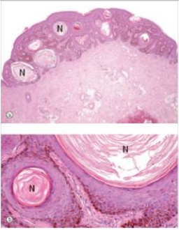
A)Actinic keratosis
B)Basal cell carcinoma
C)Compound naevus
D)Malignant melanoma
E)Seborrhoeic keratosis
F)Superficial spreading malignant melanoma
G)Squamous cell carcinoma
A)Actinic keratosis
B)Basal cell carcinoma
C)Compound naevus
D)Malignant melanoma
E)Seborrhoeic keratosis
F)Superficial spreading malignant melanoma
G)Squamous cell carcinoma
Question
A 35 year old woman had silver patches surrounded by erythema (redness)on her elbows and knees.A biopsy was done,and a representative section is shown in the accompanying photomicrograph.Which one of the following is the best diagnosis? 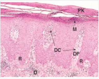
A)Basal cell carcinoma
B)Lichen planus
C)Molluscum contagiousum
D)Psoriasis
E)Squamous cell carcinoma
F)Viral warts
A)Basal cell carcinoma
B)Lichen planus
C)Molluscum contagiousum
D)Psoriasis
E)Squamous cell carcinoma
F)Viral warts
Question
A 4 year old boy developed raised bumps on his fingers.His friends at his pre-school had similar lesions.A biopsy of one of the lesions is shown in the accompanying photomicrograph.Which one of the following is the best diagnosis? 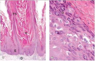
A)Basal cell carcinoma
B)Lichen planus
C)Molluscum contagiousum
D)Psoriasis
E)Squamous cell carcinoma
F)Viral warts
A)Basal cell carcinoma
B)Lichen planus
C)Molluscum contagiousum
D)Psoriasis
E)Squamous cell carcinoma
F)Viral warts
Question
A 39 year old female consulted her GP because of a small,non-healing ulcer that appeared on her nose.Upon examination,she was overly tanned,and had a 0.8 cm diameter lesion with pearly white raised edges on the right side of her nose.The lesion was completely excised.A representative section is shown in the accompanying photomicrograph.Which one of the following is the best diagnosis? 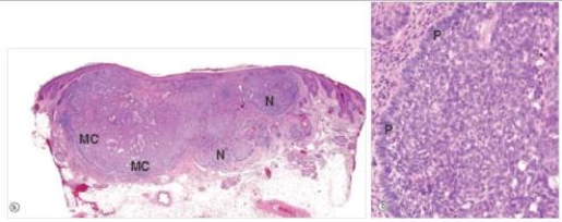
A)Actinic keratosis
B)Basal cell carcinoma
C)Compound naevus
D)Malignant melanoma
E)Seborrheic keratosis
F)Superficial spreading malignant melanoma
G)Squamous cell carcinoma
A)Actinic keratosis
B)Basal cell carcinoma
C)Compound naevus
D)Malignant melanoma
E)Seborrheic keratosis
F)Superficial spreading malignant melanoma
G)Squamous cell carcinoma
Question
How would you classify the pigmented lesion shown in the accompanying photomicrograph? 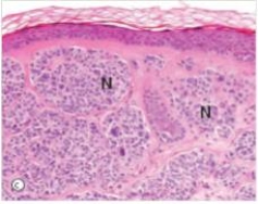
A)Intradermal naevus
B)Junctional naevus
C)Malignant melanoma
D)Seborrheic keratosis
E)Superficial spreading melanoma
A)Intradermal naevus
B)Junctional naevus
C)Malignant melanoma
D)Seborrheic keratosis
E)Superficial spreading melanoma
Question
A 4 year old boy developed raised bumps on his fingers.His friends at his pre-school had similar lesions.A biopsy of one of the lesions is shown in the accompanying photomicrograph.Which one of the following is the best diagnosis? 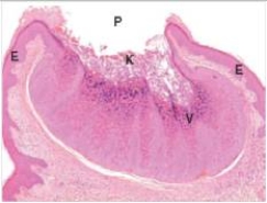
A)Basal cell carcinoma
B)Lichen planus
C)Molluscum contagiousum
D)Psoriasis
E)Squamous cell carcinoma
F)Viral warts
A)Basal cell carcinoma
B)Lichen planus
C)Molluscum contagiousum
D)Psoriasis
E)Squamous cell carcinoma
F)Viral warts
Question
A 45 year old woman noticed a whitish patch on the skin of her forearm.Her GP biopsied the lesion,which a pathologist eventually diagnosed as lichen planus.A representative section is shown in the accompanying photomicrograph.How would you characterise the structure marked "CB" in the high power photo on the right?
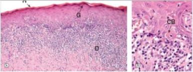
A)Apoptotic body
B)Artifact
C)Fungus
D)Mite
E)Inflammatory cell
A)Apoptotic body
B)Artifact
C)Fungus
D)Mite
E)Inflammatory cell
Question
The thickening of the epidermis in this photomicrograph is due primarily to enlargement of which one of the following epidermal layers? 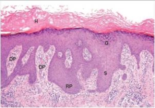
A)Dermis
B)Dermal papillae
C)Keratin layer (hyperkeratosis)
D)Rete ridges (acanthosis)
A)Dermis
B)Dermal papillae
C)Keratin layer (hyperkeratosis)
D)Rete ridges (acanthosis)

Unlock Deck
Sign up to unlock the cards in this deck!
Unlock Deck
Unlock Deck
1/8
Play
Full screen (f)
Deck 21: Skin
1
A 75 year old man had several brown,raised lesions on his arms.One of them began to bleed,after he snagged it while putting on his shirt.Fearing melanoma,he consulted his GP,who removed it.A representative section is shown in the accompanying photomicrograph.Which one of the following is the best diagnosis?
A)Actinic keratosis
B)Basal cell carcinoma
C)Compound naevus
D)Malignant melanoma
E)Seborrhoeic keratosis
F)Superficial spreading malignant melanoma
G)Squamous cell carcinoma
A)Actinic keratosis
B)Basal cell carcinoma
C)Compound naevus
D)Malignant melanoma
E)Seborrhoeic keratosis
F)Superficial spreading malignant melanoma
G)Squamous cell carcinoma
Seborrhoeic keratosis
2
A 35 year old woman had silver patches surrounded by erythema (redness)on her elbows and knees.A biopsy was done,and a representative section is shown in the accompanying photomicrograph.Which one of the following is the best diagnosis?
A)Basal cell carcinoma
B)Lichen planus
C)Molluscum contagiousum
D)Psoriasis
E)Squamous cell carcinoma
F)Viral warts
A)Basal cell carcinoma
B)Lichen planus
C)Molluscum contagiousum
D)Psoriasis
E)Squamous cell carcinoma
F)Viral warts
Psoriasis
3
A 4 year old boy developed raised bumps on his fingers.His friends at his pre-school had similar lesions.A biopsy of one of the lesions is shown in the accompanying photomicrograph.Which one of the following is the best diagnosis?
A)Basal cell carcinoma
B)Lichen planus
C)Molluscum contagiousum
D)Psoriasis
E)Squamous cell carcinoma
F)Viral warts
A)Basal cell carcinoma
B)Lichen planus
C)Molluscum contagiousum
D)Psoriasis
E)Squamous cell carcinoma
F)Viral warts
Viral warts
4
A 39 year old female consulted her GP because of a small,non-healing ulcer that appeared on her nose.Upon examination,she was overly tanned,and had a 0.8 cm diameter lesion with pearly white raised edges on the right side of her nose.The lesion was completely excised.A representative section is shown in the accompanying photomicrograph.Which one of the following is the best diagnosis?
A)Actinic keratosis
B)Basal cell carcinoma
C)Compound naevus
D)Malignant melanoma
E)Seborrheic keratosis
F)Superficial spreading malignant melanoma
G)Squamous cell carcinoma
A)Actinic keratosis
B)Basal cell carcinoma
C)Compound naevus
D)Malignant melanoma
E)Seborrheic keratosis
F)Superficial spreading malignant melanoma
G)Squamous cell carcinoma
Unlock Deck
Unlock for access to all 8 flashcards in this deck.
Unlock Deck
k this deck
5
How would you classify the pigmented lesion shown in the accompanying photomicrograph?
A)Intradermal naevus
B)Junctional naevus
C)Malignant melanoma
D)Seborrheic keratosis
E)Superficial spreading melanoma
A)Intradermal naevus
B)Junctional naevus
C)Malignant melanoma
D)Seborrheic keratosis
E)Superficial spreading melanoma
Unlock Deck
Unlock for access to all 8 flashcards in this deck.
Unlock Deck
k this deck
6
A 4 year old boy developed raised bumps on his fingers.His friends at his pre-school had similar lesions.A biopsy of one of the lesions is shown in the accompanying photomicrograph.Which one of the following is the best diagnosis?
A)Basal cell carcinoma
B)Lichen planus
C)Molluscum contagiousum
D)Psoriasis
E)Squamous cell carcinoma
F)Viral warts
A)Basal cell carcinoma
B)Lichen planus
C)Molluscum contagiousum
D)Psoriasis
E)Squamous cell carcinoma
F)Viral warts
Unlock Deck
Unlock for access to all 8 flashcards in this deck.
Unlock Deck
k this deck
7
A 45 year old woman noticed a whitish patch on the skin of her forearm.Her GP biopsied the lesion,which a pathologist eventually diagnosed as lichen planus.A representative section is shown in the accompanying photomicrograph.How would you characterise the structure marked "CB" in the high power photo on the right?
A)Apoptotic body
B)Artifact
C)Fungus
D)Mite
E)Inflammatory cell
A)Apoptotic body
B)Artifact
C)Fungus
D)Mite
E)Inflammatory cell
Unlock Deck
Unlock for access to all 8 flashcards in this deck.
Unlock Deck
k this deck
8
The thickening of the epidermis in this photomicrograph is due primarily to enlargement of which one of the following epidermal layers?
A)Dermis
B)Dermal papillae
C)Keratin layer (hyperkeratosis)
D)Rete ridges (acanthosis)
A)Dermis
B)Dermal papillae
C)Keratin layer (hyperkeratosis)
D)Rete ridges (acanthosis)
Unlock Deck
Unlock for access to all 8 flashcards in this deck.
Unlock Deck
k this deck
Unlock Deck
Unlock for access to all 8 flashcards in this deck.


