Deck 13: Gastrointestinal System
Full screen (f)
Question
A 26 year old medical student reported epigastric pain and discomfort,often around exam time.Upper GI endoscopy was performed,which reveal oedematous,reddish gastric mucosa.A biopsy was done,a representative section of which is shown in the accompanying photomicrograph.The right panel shows a Cresyl violet special stain.Which one of the following is the best diagnosis? 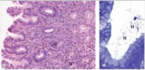
A)Gastric adenocarcinoma
B)Giardia lamblia
C)Helicobacter pylori gastritis
D)Peptic ulcer
E)Psychosomatic "tummy ache"
A)Gastric adenocarcinoma
B)Giardia lamblia
C)Helicobacter pylori gastritis
D)Peptic ulcer
E)Psychosomatic "tummy ache"
Question
A 35 year old mechanic was experiencing excruciating bouts of bloody diarrhoea.He consulted his local physician,who recommended partial colectomy in order to control the bleeding.A representative section of the specimen is shown in the accompanying low power photomicrograph.Which one of the following is the most likely diagnosis? 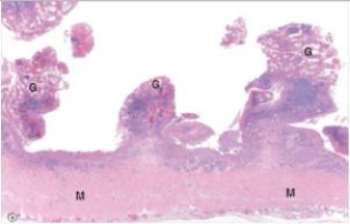
A)Adenocarcinoma
B)Crohn's disease
C)Giardia lamblia
D)Peptic ulcer disease
E)Ulcerative colitis
A)Adenocarcinoma
B)Crohn's disease
C)Giardia lamblia
D)Peptic ulcer disease
E)Ulcerative colitis
Question
A 48 year old Japanese man who had recently immigrated to the United States was seen by his local physician for fatigue,weight loss,and "feeling full all the time".Endoscopy revealed an absence of rugal folds,and the stomach was difficult to distend with air,so-called "leather bottle stomach".A biopsy was performed,and a representative section is shown in the accompanying photomicrograph.Which one of the following is the best diagnosis? 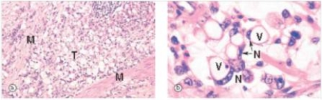
A)Amoebiasis
B)Gastric carcinoma
C)Granulomatous inflammation
D)Peptic ulcer
E)Signet ring cell gastric carcinoma,diffuse type
A)Amoebiasis
B)Gastric carcinoma
C)Granulomatous inflammation
D)Peptic ulcer
E)Signet ring cell gastric carcinoma,diffuse type
Question
A 24 year old nursing student was having difficulty keeping up in class because of extreme fatigue.She sought help from the Students' Health Service.A full blood count (FBC)showed anaemia with increased RBC (red blood cell)size (macrocytic anaemia).An upper GI endoscopy was performed,and biopsies taken.A representative section of the biopsy is shown in the accompanying photomicrograph,with normal small bowel shown on the left for comparison (villi are identified as "V").Which one of the following is the best diagnosis? 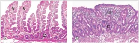
A)Adenocarcinoma
B)Coeliac disease
C)Crohn's disease
D)Iron deficiency anemia
E)Ulcerative colitis
A)Adenocarcinoma
B)Coeliac disease
C)Crohn's disease
D)Iron deficiency anemia
E)Ulcerative colitis
Question
A 48 year old Japanese man who had recently immigrated to the United States was seen by his local physician for fatigue,weight loss,and "feeling full all the time".Endoscopy revealed a polypoid tumour mass with "heaped up" edges.A biopsy was performed,and a representative section is shown in the accompanying photomicrograph.Which one of the following is the best diagnosis? 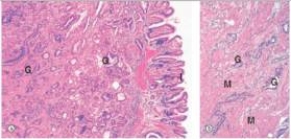
A)Gastric adenocarcinoma
B)Giardia lamblia
C)Helicobacter pylori gastritis
D)Peptic ulcer
E)Signet ring carcinoma
A)Gastric adenocarcinoma
B)Giardia lamblia
C)Helicobacter pylori gastritis
D)Peptic ulcer
E)Signet ring carcinoma
Question
A 25 year old woman woke one morning feeling something wet and warm on her abdomen.When she pulled back the covers,she discovered it was liquid faeces,leaking from a fistulous opening on her abdominal wall,which was soon discovered to have originated in her ileum.She recounted that she had been experiencing diarrhoea for several weeks prior to this event,but had not sought medical attention.At the hospital,a section of small bowel was resected,and the fistula repaired.A representative section of the surgical specimen is shown in the accompanying photomicrograph,with a high power view on the right.Which one of the following is the best diagnosis? 
A)Adenocarcinoma
B)Crohn's disease
C)Peptic ulcer
D)Tuberculosis
E)Ulcerative colitis
A)Adenocarcinoma
B)Crohn's disease
C)Peptic ulcer
D)Tuberculosis
E)Ulcerative colitis
Question
A 48 year old man became alarmed when he noted some bright red blood in his stool.He consulted a local physician,who palpated an indurated area (firm to touch)in the patient's rectum.Surgery was performed,and a representative section of the surgical specimen is show on low power in the accompanying photomicrograph.Which one of the following is the best diagnosis? 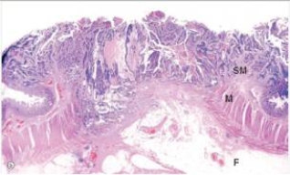
A)Adenomatous polyp
B)Invasive adenocarcinoma
C)Tubulovillous adenoma
D)Ulcerative colitis
E)Villous adenoma
A)Adenomatous polyp
B)Invasive adenocarcinoma
C)Tubulovillous adenoma
D)Ulcerative colitis
E)Villous adenoma
Question
A 38 year old male consulted his local physician because he was experiencing strange episodes of skin flushing and sweating.The physician suspected a GI tumour,and ordered a CT scan,which disclosed a mass in the duodenum,and possible metastatic disease in the liver.The duodenal mass was biopsied,and is shown in the accompanying high power photomicrograph.An immunohistochemistry stain for chromogranin is shown on the right.Which one of the following is the best diagnosis? 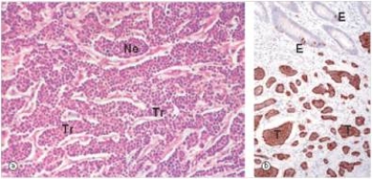
A)Adenocarcinoma
B)Carcinoid tumour
C)Small cell carcinoma
D)Pancreatic carcinoma
E)Transitional cell carcinoma
A)Adenocarcinoma
B)Carcinoid tumour
C)Small cell carcinoma
D)Pancreatic carcinoma
E)Transitional cell carcinoma

Unlock Deck
Sign up to unlock the cards in this deck!
Unlock Deck
Unlock Deck
1/8
Play
Full screen (f)
Deck 13: Gastrointestinal System
1
A 26 year old medical student reported epigastric pain and discomfort,often around exam time.Upper GI endoscopy was performed,which reveal oedematous,reddish gastric mucosa.A biopsy was done,a representative section of which is shown in the accompanying photomicrograph.The right panel shows a Cresyl violet special stain.Which one of the following is the best diagnosis?
A)Gastric adenocarcinoma
B)Giardia lamblia
C)Helicobacter pylori gastritis
D)Peptic ulcer
E)Psychosomatic "tummy ache"
A)Gastric adenocarcinoma
B)Giardia lamblia
C)Helicobacter pylori gastritis
D)Peptic ulcer
E)Psychosomatic "tummy ache"
Helicobacter pylori gastritis
2
A 35 year old mechanic was experiencing excruciating bouts of bloody diarrhoea.He consulted his local physician,who recommended partial colectomy in order to control the bleeding.A representative section of the specimen is shown in the accompanying low power photomicrograph.Which one of the following is the most likely diagnosis?
A)Adenocarcinoma
B)Crohn's disease
C)Giardia lamblia
D)Peptic ulcer disease
E)Ulcerative colitis
A)Adenocarcinoma
B)Crohn's disease
C)Giardia lamblia
D)Peptic ulcer disease
E)Ulcerative colitis
Ulcerative colitis
3
A 48 year old Japanese man who had recently immigrated to the United States was seen by his local physician for fatigue,weight loss,and "feeling full all the time".Endoscopy revealed an absence of rugal folds,and the stomach was difficult to distend with air,so-called "leather bottle stomach".A biopsy was performed,and a representative section is shown in the accompanying photomicrograph.Which one of the following is the best diagnosis?
A)Amoebiasis
B)Gastric carcinoma
C)Granulomatous inflammation
D)Peptic ulcer
E)Signet ring cell gastric carcinoma,diffuse type
A)Amoebiasis
B)Gastric carcinoma
C)Granulomatous inflammation
D)Peptic ulcer
E)Signet ring cell gastric carcinoma,diffuse type
Signet ring cell gastric carcinoma,diffuse type
4
A 24 year old nursing student was having difficulty keeping up in class because of extreme fatigue.She sought help from the Students' Health Service.A full blood count (FBC)showed anaemia with increased RBC (red blood cell)size (macrocytic anaemia).An upper GI endoscopy was performed,and biopsies taken.A representative section of the biopsy is shown in the accompanying photomicrograph,with normal small bowel shown on the left for comparison (villi are identified as "V").Which one of the following is the best diagnosis?
A)Adenocarcinoma
B)Coeliac disease
C)Crohn's disease
D)Iron deficiency anemia
E)Ulcerative colitis
A)Adenocarcinoma
B)Coeliac disease
C)Crohn's disease
D)Iron deficiency anemia
E)Ulcerative colitis
Unlock Deck
Unlock for access to all 8 flashcards in this deck.
Unlock Deck
k this deck
5
A 48 year old Japanese man who had recently immigrated to the United States was seen by his local physician for fatigue,weight loss,and "feeling full all the time".Endoscopy revealed a polypoid tumour mass with "heaped up" edges.A biopsy was performed,and a representative section is shown in the accompanying photomicrograph.Which one of the following is the best diagnosis?
A)Gastric adenocarcinoma
B)Giardia lamblia
C)Helicobacter pylori gastritis
D)Peptic ulcer
E)Signet ring carcinoma
A)Gastric adenocarcinoma
B)Giardia lamblia
C)Helicobacter pylori gastritis
D)Peptic ulcer
E)Signet ring carcinoma
Unlock Deck
Unlock for access to all 8 flashcards in this deck.
Unlock Deck
k this deck
6
A 25 year old woman woke one morning feeling something wet and warm on her abdomen.When she pulled back the covers,she discovered it was liquid faeces,leaking from a fistulous opening on her abdominal wall,which was soon discovered to have originated in her ileum.She recounted that she had been experiencing diarrhoea for several weeks prior to this event,but had not sought medical attention.At the hospital,a section of small bowel was resected,and the fistula repaired.A representative section of the surgical specimen is shown in the accompanying photomicrograph,with a high power view on the right.Which one of the following is the best diagnosis?
A)Adenocarcinoma
B)Crohn's disease
C)Peptic ulcer
D)Tuberculosis
E)Ulcerative colitis
A)Adenocarcinoma
B)Crohn's disease
C)Peptic ulcer
D)Tuberculosis
E)Ulcerative colitis
Unlock Deck
Unlock for access to all 8 flashcards in this deck.
Unlock Deck
k this deck
7
A 48 year old man became alarmed when he noted some bright red blood in his stool.He consulted a local physician,who palpated an indurated area (firm to touch)in the patient's rectum.Surgery was performed,and a representative section of the surgical specimen is show on low power in the accompanying photomicrograph.Which one of the following is the best diagnosis?
A)Adenomatous polyp
B)Invasive adenocarcinoma
C)Tubulovillous adenoma
D)Ulcerative colitis
E)Villous adenoma
A)Adenomatous polyp
B)Invasive adenocarcinoma
C)Tubulovillous adenoma
D)Ulcerative colitis
E)Villous adenoma
Unlock Deck
Unlock for access to all 8 flashcards in this deck.
Unlock Deck
k this deck
8
A 38 year old male consulted his local physician because he was experiencing strange episodes of skin flushing and sweating.The physician suspected a GI tumour,and ordered a CT scan,which disclosed a mass in the duodenum,and possible metastatic disease in the liver.The duodenal mass was biopsied,and is shown in the accompanying high power photomicrograph.An immunohistochemistry stain for chromogranin is shown on the right.Which one of the following is the best diagnosis?
A)Adenocarcinoma
B)Carcinoid tumour
C)Small cell carcinoma
D)Pancreatic carcinoma
E)Transitional cell carcinoma
A)Adenocarcinoma
B)Carcinoid tumour
C)Small cell carcinoma
D)Pancreatic carcinoma
E)Transitional cell carcinoma
Unlock Deck
Unlock for access to all 8 flashcards in this deck.
Unlock Deck
k this deck
Unlock Deck
Unlock for access to all 8 flashcards in this deck.


