Deck 22: Heart
Full screen (f)
Question
Question
Question
Question
Question
Question
Question
Question
Question
Question
Question
Question
Question
Question
Question
Question
Question
Question
Question
Question
Question
Question
Question
Question
Question
Question
Question
Question
Question
Question
Question
Question
Question
Question
Question
Question
Question
Question
Question
Question
Question
Question
Question
Question
Question
Question
Question
Question
Question
Question
Question
Question
Question
Question
Question
Question
Question
Question
Question
Question
Question
Question
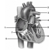
In this figure showing an anterior view of the heart,the left atrium is noted by number _____.
A)1
B)2
C)3
D)4
E)5
Question
Question
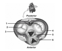
In this figure showing an oblique section of the heart,the pulmonary semilunar valve is number
A)1.
B)2.
C)3.
D)5.
E)6.
Question
Question
Question
Question
Question
Question
Question
Question
Question
In this figure showing an anterior view of the heart,number 7 depicts the
A)aortic semilunar valve.
B)right atrium.
C)left ventricle.
D)right atrioventricular valve.
E)pulmonary semilunar valve.
Question
Question
In this figure showing an oblique section of the heart,number 3 depicts the
A)wall of the left ventricle.
B)wall of the right atrium.
C)wall of the left atrium.
D)wall of the right ventricle.
E)left atrioventricular valve.
Question
Question
Question
Question
Question

Unlock Deck
Sign up to unlock the cards in this deck!
Unlock Deck
Unlock Deck
1/101
Play
Full screen (f)
Deck 22: Heart
1
Which heart chambers pump deoxygenated blood?
A)Left atrium and right atrium
B)Left ventricle and right ventricle
C)Right atrium and right ventricle
D)Left atrium and left ventricle
E)Right atrium and left ventricle
A)Left atrium and right atrium
B)Left ventricle and right ventricle
C)Right atrium and right ventricle
D)Left atrium and left ventricle
E)Right atrium and left ventricle
C
2
The epicardium is another name for the
A)visceral layer of the serous pericardium.
B)parietal layer of the serous pericardium.
C)pericardium.
D)myocardium.
E)myometrium.
A)visceral layer of the serous pericardium.
B)parietal layer of the serous pericardium.
C)pericardium.
D)myocardium.
E)myometrium.
A
3
The cell-to-cell contacts of the cardiac muscle fibers are called
A)Z discs.
B)T-tubules.
C)intercalated discs.
D)cardiac discs.
E)sarcoplasmic reticulum.
A)Z discs.
B)T-tubules.
C)intercalated discs.
D)cardiac discs.
E)sarcoplasmic reticulum.
C
4
Which pair is located more anteriorly in a heart in normal position?
A)Left atrium and left ventricle
B)Left atrium and right ventricle
C)Right atrium and left atrium
D)Right atrium and right ventricle
E)Right atrium and left ventricle
A)Left atrium and left ventricle
B)Left atrium and right ventricle
C)Right atrium and left atrium
D)Right atrium and right ventricle
E)Right atrium and left ventricle
Unlock Deck
Unlock for access to all 101 flashcards in this deck.
Unlock Deck
k this deck
5
It is the _____________ that permits the compression necessary to pump large volumes of blood out of the ventricles.
A)negative pressure inside the ventricles
B)absence of oxygenated blood in the atria
C)arrangement of cardiac muscle in the heart wall
D)presence of skeletal muscle tissue in the heart skeleton
E)presence of papillary muscles in the ventricles
A)negative pressure inside the ventricles
B)absence of oxygenated blood in the atria
C)arrangement of cardiac muscle in the heart wall
D)presence of skeletal muscle tissue in the heart skeleton
E)presence of papillary muscles in the ventricles
Unlock Deck
Unlock for access to all 101 flashcards in this deck.
Unlock Deck
k this deck
6
The left atrium and left ventricle are part of which circuit of the cardiovascular system?
A)Pulmonary circuit
B)Cardio circuit
C)Coronary circuit
D)Systemic circuit
E)Schwann circuit
A)Pulmonary circuit
B)Cardio circuit
C)Coronary circuit
D)Systemic circuit
E)Schwann circuit
Unlock Deck
Unlock for access to all 101 flashcards in this deck.
Unlock Deck
k this deck
7
Which can be used to characterize blood flow in the human body?
A: There is a unidirectional blood flow.
B: Arteries always carry oxygenated blood.
C: Veins always carry deoxygenated blood.
D: Arteries carry blood away from the heart.
E: Veins carry blood toward the heart.
A)a,c,d,e
B)a,b,c,d,e
C)a,d,e
D)b,c,d,e
E)d,e
A: There is a unidirectional blood flow.
B: Arteries always carry oxygenated blood.
C: Veins always carry deoxygenated blood.
D: Arteries carry blood away from the heart.
E: Veins carry blood toward the heart.
A)a,c,d,e
B)a,b,c,d,e
C)a,d,e
D)b,c,d,e
E)d,e
Unlock Deck
Unlock for access to all 101 flashcards in this deck.
Unlock Deck
k this deck
8
Which analogy fits the human heart?
A)It is like a single pump.
B)It is like a double pump,each working side by side with the other.
C)It is like four pumps working in alternating cycles.
D)It is like a double pump,each working at its own rate determined by the needs of the body served.
E)It is like a single pump whose various chambers all work together at once.
Unlock Deck
Unlock for access to all 101 flashcards in this deck.
Unlock Deck
k this deck
9
Which circuit of the cardiovascular system is responsible for sending blood to the kidneys,stomach,and pelvic regions?
A)Pulmonary circuit
B)Innards circuit
C)Vagus circuit
D)Systemic circuit
E)Vanguard circuit
A)Pulmonary circuit
B)Innards circuit
C)Vagus circuit
D)Systemic circuit
E)Vanguard circuit
Unlock Deck
Unlock for access to all 101 flashcards in this deck.
Unlock Deck
k this deck
10
The inferior chambers of the heart are the
A)atria.
B)auricles.
C)ventricles.
D)sulci.
E)gyri.
A)atria.
B)auricles.
C)ventricles.
D)sulci.
E)gyri.
Unlock Deck
Unlock for access to all 101 flashcards in this deck.
Unlock Deck
k this deck
11
The base of the heart is formed primarily by the
A)right atrium.
B)right ventricle.
C)left atrium.
D)left ventricle.
E)None of these choices is correct.
A)right atrium.
B)right ventricle.
C)left atrium.
D)left ventricle.
E)None of these choices is correct.
Unlock Deck
Unlock for access to all 101 flashcards in this deck.
Unlock Deck
k this deck
12
Which correctly describes the heart's apex?
A)Projects slightly anteroinferiorly toward the left side of the body
B)Projects slightly anteroinferiorly toward the right side of the body
C)Projects slightly posteroinferiorly toward the left side of the body
D)Projects slightly posteroinferiorly toward the right side of the body
E)Projects slightly posteroinferiorly toward the midline of the body
Unlock Deck
Unlock for access to all 101 flashcards in this deck.
Unlock Deck
k this deck
13
Which are differences between cardiac muscle tissue and skeletal muscle tissue?
A: The sarcoplasmic reticulum in cardiac muscle is less extensive.
B: The sarcoplasmic reticulum in cardiac muscle is more organized.
C: Cardiac muscle has intercalated discs;skeletal muscle does not.
D: Cardiac muscle has 1 or 2 nuclei per cell;skeletal muscle has multiple nuclei per cell.
E: Cardiac muscle has more well defined terminal cisternae.
A)a,c,d
B)a,c,e
C)b,c,e
D)a,b,e
E)b,d,e
Unlock Deck
Unlock for access to all 101 flashcards in this deck.
Unlock Deck
k this deck
14
Cardiac muscle fibers
A)contract as a single unit.
B)are only loosely connected by the intercalated discs.
C)have a low oxygen need.
D)utilize hemoglobin as an energy source.
A)contract as a single unit.
B)are only loosely connected by the intercalated discs.
C)have a low oxygen need.
D)utilize hemoglobin as an energy source.
Unlock Deck
Unlock for access to all 101 flashcards in this deck.
Unlock Deck
k this deck
15
The serous fluid within the pericardial cavity works to
A)lubricate the membranes of the serous pericardium.
B)slow the heart rate.
C)equalize the pressure in the great vessels.
D)eliminate blood pressure spikes.
A)lubricate the membranes of the serous pericardium.
B)slow the heart rate.
C)equalize the pressure in the great vessels.
D)eliminate blood pressure spikes.
Unlock Deck
Unlock for access to all 101 flashcards in this deck.
Unlock Deck
k this deck
16
The layer of the heart wall composed of cardiac muscle tissue is the
A)pericardium.
B)myocardium.
C)endocardium.
D)pericardial cavity.
E)pectinate muscle.
A)pericardium.
B)myocardium.
C)endocardium.
D)pericardial cavity.
E)pectinate muscle.
Unlock Deck
Unlock for access to all 101 flashcards in this deck.
Unlock Deck
k this deck
17
Which values are reasonable for a healthy,80 kilogram resting adult?
A)5.25 liters of blood pumped per ventricle per minute;108,000 beats per day
B)4.25 liters of blood pumped per ventricle per minute;60,000 beats per day
C)6.75 liters of blood pumped per ventricle per minute;20,000 beats per day
D)7.25 liters of blood pumped per ventricle per minute;76,000 beats per day
E)5.75 liters of blood pumped per ventricle per minute;144,000 beats per day
Unlock Deck
Unlock for access to all 101 flashcards in this deck.
Unlock Deck
k this deck
18
Which of the cardiovascular system's circuits has deoxygenated blood in its arteries?
A)Pulmonary circuit
B)Innards circuit
C)Antithesis circuit
D)Systemic circuit
E)Vanguard circuit
A)Pulmonary circuit
B)Innards circuit
C)Antithesis circuit
D)Systemic circuit
E)Vanguard circuit
Unlock Deck
Unlock for access to all 101 flashcards in this deck.
Unlock Deck
k this deck
19
Which describes the endocardium?
A: Has single layer of epithelium
B: Has layer of areolar connective tissue
C: Epithelial cells are squamous
D: Epithelial cells are cuboidal
E: Has layer of adipose connective tissue
F: Has patches of myocardium
A)a,b,c
B)a,b,d
C)a,d,e
D)a,b,c,e
E)a,e,f
A: Has single layer of epithelium
B: Has layer of areolar connective tissue
C: Epithelial cells are squamous
D: Epithelial cells are cuboidal
E: Has layer of adipose connective tissue
F: Has patches of myocardium
A)a,b,c
B)a,b,d
C)a,d,e
D)a,b,c,e
E)a,e,f
Unlock Deck
Unlock for access to all 101 flashcards in this deck.
Unlock Deck
k this deck
20
The arteries and veins that leave and enter the heart are called the great vessels because
A)they are associated with the heart directly.
B)they carry the most highly oxygenated blood.
C)they are longer than any other blood vessels in the body.
D)they have relatively large diameters.
E)Both types have the highest blood pressure in the body.
A)they are associated with the heart directly.
B)they carry the most highly oxygenated blood.
C)they are longer than any other blood vessels in the body.
D)they have relatively large diameters.
E)Both types have the highest blood pressure in the body.
Unlock Deck
Unlock for access to all 101 flashcards in this deck.
Unlock Deck
k this deck
21
The beginning of the cardiac cycle is when the
A)atria fill with blood.
B)ventricles fill with blood.
C)atria contract.
D)two semilunar valves close.
E)ventricles contract.
A)atria fill with blood.
B)ventricles fill with blood.
C)atria contract.
D)two semilunar valves close.
E)ventricles contract.
Unlock Deck
Unlock for access to all 101 flashcards in this deck.
Unlock Deck
k this deck
22
The function of the coronary sinus is to
A)connect the top and bottom halves of the heart.
B)guide the aorta out of the heart.
C)guide the inferior vena cava into the right atrium.
D)take blood from the coronary circulation to the right atrium.
E)shunt blood from the right atrium to the left atrium.
A)connect the top and bottom halves of the heart.
B)guide the aorta out of the heart.
C)guide the inferior vena cava into the right atrium.
D)take blood from the coronary circulation to the right atrium.
E)shunt blood from the right atrium to the left atrium.
Unlock Deck
Unlock for access to all 101 flashcards in this deck.
Unlock Deck
k this deck
23
The atria are separated from the ventricles externally by the
A)anterior interventricular sulcus.
B)posterior interventricular sulcus.
C)sinoventricular sulcus.
D)coronary sulcus.
A)anterior interventricular sulcus.
B)posterior interventricular sulcus.
C)sinoventricular sulcus.
D)coronary sulcus.
Unlock Deck
Unlock for access to all 101 flashcards in this deck.
Unlock Deck
k this deck
24
The heart valves
A)stabilize and hold the arteries leaving the heart.
B)permit the passage of blood in one direction.
C)separate the right and left sides of the heart.
D)are only used in the fetal heart.
E)direct the conduction impulse through the heart muscle.
A)stabilize and hold the arteries leaving the heart.
B)permit the passage of blood in one direction.
C)separate the right and left sides of the heart.
D)are only used in the fetal heart.
E)direct the conduction impulse through the heart muscle.
Unlock Deck
Unlock for access to all 101 flashcards in this deck.
Unlock Deck
k this deck
25
As blood is pumped into the arterial trunks past the semilunar valves,it
A)pushes against the cusps,forcing the valves open.
B)pushes against the cusps,forcing the valves closed.
C)fills the cusps,causing them to expand and block the backflow of blood.
D)fills the cusps,causing them to expand and open up for the flow of blood.
E)None of these answers is correct.
A)pushes against the cusps,forcing the valves open.
B)pushes against the cusps,forcing the valves closed.
C)fills the cusps,causing them to expand and block the backflow of blood.
D)fills the cusps,causing them to expand and open up for the flow of blood.
E)None of these answers is correct.
Unlock Deck
Unlock for access to all 101 flashcards in this deck.
Unlock Deck
k this deck
26
Which structure marks the end of the left ventricle and the entrance to the aorta?
A)Pulmonary semilunar valve
B)Left atrioventricular valve
C)Right atrioventricular valve
D)Aortic semilunar valve
E)Foramen ovale
A)Pulmonary semilunar valve
B)Left atrioventricular valve
C)Right atrioventricular valve
D)Aortic semilunar valve
E)Foramen ovale
Unlock Deck
Unlock for access to all 101 flashcards in this deck.
Unlock Deck
k this deck
27
Which are functions performed by the fibrous skeleton of the heart?
A: Separates the atria and ventricles
B: Anchors the heart valves
C: Provides electrical insulation between the atria and ventricles
D: Provides the framework for the attachment of the myocardium
E: None of these are true functions of the fibrous skeleton of the heart.
A)a,d
B)b,d
C)a,c,d
D)a,b,c,d
E)e
Unlock Deck
Unlock for access to all 101 flashcards in this deck.
Unlock Deck
k this deck
28
The papillary muscles attach to the cusps of the atrioventricular valves by means of the
A)pectinate muscles.
B)trabeculae carneae.
C)conus arteriosus.
D)chordae tendineae.
E)tricuspid valve.
A)pectinate muscles.
B)trabeculae carneae.
C)conus arteriosus.
D)chordae tendineae.
E)tricuspid valve.
Unlock Deck
Unlock for access to all 101 flashcards in this deck.
Unlock Deck
k this deck
29
How many half-moon shaped,pocketlike cusps are found in each semilunar valve?
A)1
B)2
C)3
D)4
E)6
A)1
B)2
C)3
D)4
E)6
Unlock Deck
Unlock for access to all 101 flashcards in this deck.
Unlock Deck
k this deck
30
Typically,there are __________ papillary muscles that project from the wall of the left ventricle and attach to the chordae tendineae that support the left AV valve.
A)three
B)two
C)a highly variable number of
D)six
E)no
A)three
B)two
C)a highly variable number of
D)six
E)no
Unlock Deck
Unlock for access to all 101 flashcards in this deck.
Unlock Deck
k this deck
31
The internal wall surface of each ventricle displays large,smooth,irregular muscular ridges called
A)conus arteriosus.
B)atrioventricular opening.
C)trabeculae carneae.
D)chordae tendineae
E)pectinate muscles
A)conus arteriosus.
B)atrioventricular opening.
C)trabeculae carneae.
D)chordae tendineae
E)pectinate muscles
Unlock Deck
Unlock for access to all 101 flashcards in this deck.
Unlock Deck
k this deck
32
During a cardiac cycle,how many of the four chambers contract at any one time?
A)1
B)2
C)4
D)Highly variable,depending on the heart beat rate
E)None of these answers is correct.
A)1
B)2
C)4
D)Highly variable,depending on the heart beat rate
E)None of these answers is correct.
Unlock Deck
Unlock for access to all 101 flashcards in this deck.
Unlock Deck
k this deck
33
Which bears a close structural relationship with the fossa ovalis?
A)Papillary muscles
B)Trabeculae carneae
C)Conus arteriosus
D)Chordae tendineae
E)Foramen ovale
A)Papillary muscles
B)Trabeculae carneae
C)Conus arteriosus
D)Chordae tendineae
E)Foramen ovale
Unlock Deck
Unlock for access to all 101 flashcards in this deck.
Unlock Deck
k this deck
34
Pectinate muscles are found on the
A)anterior wall of the right atrium.
B)posterior wall of the right ventricle.
C)anterior wall of the right ventricle.
D)anterior wall of the right and left atria.
E)posterior wall of the right and left ventricles.
A)anterior wall of the right atrium.
B)posterior wall of the right ventricle.
C)anterior wall of the right ventricle.
D)anterior wall of the right and left atria.
E)posterior wall of the right and left ventricles.
Unlock Deck
Unlock for access to all 101 flashcards in this deck.
Unlock Deck
k this deck
35
Which carries oxygenated blood from the lungs to the heart?
A)Pulmonary arteries
B)Pulmonary veins
C)Pulmonary trunk
D)Inferior vena cava
E)Superior vena cava
A)Pulmonary arteries
B)Pulmonary veins
C)Pulmonary trunk
D)Inferior vena cava
E)Superior vena cava
Unlock Deck
Unlock for access to all 101 flashcards in this deck.
Unlock Deck
k this deck
36
The trabeculae carneae in the left ventricle are ____________ in the right ventricle.
A)less prominent than
B)not present as
C)more prominent than
D)equally as prominent as
E)None of these answers is correct.
A)less prominent than
B)not present as
C)more prominent than
D)equally as prominent as
E)None of these answers is correct.
Unlock Deck
Unlock for access to all 101 flashcards in this deck.
Unlock Deck
k this deck
37
Which action causes the closure of the right atrioventricular valve?
A)Contraction of the right atrium
B)Contraction of the left atrium
C)Contraction of the right ventricle
D)Contraction of the left ventricle
E)None of these answers is correct.
A)Contraction of the right atrium
B)Contraction of the left atrium
C)Contraction of the right ventricle
D)Contraction of the left ventricle
E)None of these answers is correct.
Unlock Deck
Unlock for access to all 101 flashcards in this deck.
Unlock Deck
k this deck
38
For ___________ of the cardiac cycle,all four chambers are in diastole together.
A)none
B)one-third
C)one-quarter
D)one-half
E)three-quarters
A)none
B)one-third
C)one-quarter
D)one-half
E)three-quarters
Unlock Deck
Unlock for access to all 101 flashcards in this deck.
Unlock Deck
k this deck
39
Which valve prevents the backflow of blood into the left ventricle when the ventricles relax?
A)Left atrioventricular valve
B)Aortic semilunar valve
C)Right atrioventricular valve
D)Pulmonary semilunar valve
E)
None of these answers is correct.
A)Left atrioventricular valve
B)Aortic semilunar valve
C)Right atrioventricular valve
D)Pulmonary semilunar valve
E)
None of these answers is correct.
Unlock Deck
Unlock for access to all 101 flashcards in this deck.
Unlock Deck
k this deck
40
The chordae tendineae are made out of
A)collagen fibers.
B)elastin fibers.
C)hyaline cartilage.
D)reticulin fibers.
E)trabeculae carneae.
A)collagen fibers.
B)elastin fibers.
C)hyaline cartilage.
D)reticulin fibers.
E)trabeculae carneae.
Unlock Deck
Unlock for access to all 101 flashcards in this deck.
Unlock Deck
k this deck
41
During early ventricular systole the atria are in
A)systole.
B)diastole.
A)systole.
B)diastole.
Unlock Deck
Unlock for access to all 101 flashcards in this deck.
Unlock Deck
k this deck
42
Development of the heart commences in the _______ week.
A)first
B)third
C)fifth
D)eighth
E)tenth
A)first
B)third
C)fifth
D)eighth
E)tenth
Unlock Deck
Unlock for access to all 101 flashcards in this deck.
Unlock Deck
k this deck
43
The sequence of events in the transmission of an impulse through the heart muscle is
A: AV node
B: AV bundle
C: SA node
D: through the atria
E: through the ventricles
F: bundle branches
G: Purkinje fibers
A)c,d,a,b,f,g,e
B)d,b,a,c,f,g,e
C)b,a,d,c,f,g,e
D)f,g,d,c,b,a,e
E)c,d,a,f,b,g,e
A: AV node
B: AV bundle
C: SA node
D: through the atria
E: through the ventricles
F: bundle branches
G: Purkinje fibers
A)c,d,a,b,f,g,e
B)d,b,a,c,f,g,e
C)b,a,d,c,f,g,e
D)f,g,d,c,b,a,e
E)c,d,a,f,b,g,e
Unlock Deck
Unlock for access to all 101 flashcards in this deck.
Unlock Deck
k this deck
44
The great cardiac vein runs alongside the
A)anterior interventricular artery.
B)posterior interventricular artery.
C)right marginal artery.
D)circumflex artery.
E)coronary sinus.
A)anterior interventricular artery.
B)posterior interventricular artery.
C)right marginal artery.
D)circumflex artery.
E)coronary sinus.
Unlock Deck
Unlock for access to all 101 flashcards in this deck.
Unlock Deck
k this deck
45
Which prenatal structure forms the ascending aorta and pulmonary trunk?
A)Truncus arteriosus
B)Sinus venosus
C)Primitive atrium
D)Primitive ventricle
E)Conus cordis
A)Truncus arteriosus
B)Sinus venosus
C)Primitive atrium
D)Primitive ventricle
E)Conus cordis
Unlock Deck
Unlock for access to all 101 flashcards in this deck.
Unlock Deck
k this deck
46
Which does not drain into the coronary sinus?
A)Great cardiac vein
B)Small cardiac vein
C)Anterior interventricular vein
D)Middle cardiac vein
E)No exceptions;all choices drain into the coronary sinus.
A)Great cardiac vein
B)Small cardiac vein
C)Anterior interventricular vein
D)Middle cardiac vein
E)No exceptions;all choices drain into the coronary sinus.
Unlock Deck
Unlock for access to all 101 flashcards in this deck.
Unlock Deck
k this deck
47
Which may predispose one to inadequate coronary blood flow?
A)Bradycardia
B)Hypertension
C)Angina pectoris
D)Tachycardia
E)None of these answers is correct.
A)Bradycardia
B)Hypertension
C)Angina pectoris
D)Tachycardia
E)None of these answers is correct.
Unlock Deck
Unlock for access to all 101 flashcards in this deck.
Unlock Deck
k this deck
48
The right border of the heart is supplied by the
A)circumflex artery.
B)posterior interventricular artery.
C)anterior interventricular artery.
D)right marginal artery.
E)great cardiac vein.
A)circumflex artery.
B)posterior interventricular artery.
C)anterior interventricular artery.
D)right marginal artery.
E)great cardiac vein.
Unlock Deck
Unlock for access to all 101 flashcards in this deck.
Unlock Deck
k this deck
49
Parasympathetic innervation of the heart occurs via
A)CN IV.
B)CN VI.
C)CN VIII.
D)CN X.
E)CN XII.
A)CN IV.
B)CN VI.
C)CN VIII.
D)CN X.
E)CN XII.
Unlock Deck
Unlock for access to all 101 flashcards in this deck.
Unlock Deck
k this deck
50
In an EKG,the P wave is generated when the
A)ventricles depolarize.
B)atria depolarize.
C)atria repolarize.
D)ventricles repolarize.
E)SA node initiates an impulse.
A)ventricles depolarize.
B)atria depolarize.
C)atria repolarize.
D)ventricles repolarize.
E)SA node initiates an impulse.
Unlock Deck
Unlock for access to all 101 flashcards in this deck.
Unlock Deck
k this deck
51
What part of the cardiac conduction system is located in the posterior wall of the right atrium,adjacent to the entrance of the superior vena cava?
A)AV bundle
B)Bundle branches
C)Purkinje fibers
D)AV node
E)SA node
A)AV bundle
B)Bundle branches
C)Purkinje fibers
D)AV node
E)SA node
Unlock Deck
Unlock for access to all 101 flashcards in this deck.
Unlock Deck
k this deck
52
The left and right coronary arteries travel within the
A)anterior interventricular sulcus.
B)posterior interventricular sulcus.
C)coronary sulcus.
D)interventricular septum.
E)interatrial septum.
A)anterior interventricular sulcus.
B)posterior interventricular sulcus.
C)coronary sulcus.
D)interventricular septum.
E)interatrial septum.
Unlock Deck
Unlock for access to all 101 flashcards in this deck.
Unlock Deck
k this deck
53
During ventricular systole
A)only the AV valves open.
B)only the AV valves close.
C)only the semilunar valves close.
D)the semilunar valves close and the AV valves open.
E)the semilunar valves open and the AV valves close.
A)only the AV valves open.
B)only the AV valves close.
C)only the semilunar valves close.
D)the semilunar valves close and the AV valves open.
E)the semilunar valves open and the AV valves close.
Unlock Deck
Unlock for access to all 101 flashcards in this deck.
Unlock Deck
k this deck
54
The cardiac cycle is
A)when the ventricles are contracting.
B)when the atria are relaxed.
C)when the atria are contracting.
D)when the ventricles are relaxed.
E)all the events involved with a single heart beat.
A)when the ventricles are contracting.
B)when the atria are relaxed.
C)when the atria are contracting.
D)when the ventricles are relaxed.
E)all the events involved with a single heart beat.
Unlock Deck
Unlock for access to all 101 flashcards in this deck.
Unlock Deck
k this deck
55
During ventricular diastole
A)only the AV valves open.
B)only the AV valves close.
C)only the semilunar valves close.
D)the semilunar valves close and the AV valves open.
E)the semilunar valves open and the AV valves close.
A)only the AV valves open.
B)only the AV valves close.
C)only the semilunar valves close.
D)the semilunar valves close and the AV valves open.
E)the semilunar valves open and the AV valves close.
Unlock Deck
Unlock for access to all 101 flashcards in this deck.
Unlock Deck
k this deck
56
One example of cardiac arrhythmia is _________,in which a rapid,repetitious movement of the ventricular muscle replaces normal contractions.
A)atrial fibrillation
B)atrial flutter
C)ventricular fibrillation
D)premature ventricular contraction
E)None of the choices is correct.
A)atrial fibrillation
B)atrial flutter
C)ventricular fibrillation
D)premature ventricular contraction
E)None of the choices is correct.
Unlock Deck
Unlock for access to all 101 flashcards in this deck.
Unlock Deck
k this deck
57
Of the four "normal" heart sounds,the initial "lubb" sound is heard when the
A)AV valves close.
B)semilunar valves close.
C)AV valves open.
D)semilunar valves open.
E)foramen ovale closes.
A)AV valves close.
B)semilunar valves close.
C)AV valves open.
D)semilunar valves open.
E)foramen ovale closes.
Unlock Deck
Unlock for access to all 101 flashcards in this deck.
Unlock Deck
k this deck
58
In an EKG,the T wave denotes
A)depolarization of the atria.
B)depolarization of the right ventricle.
C)repolarization of the ventricles.
D)closure of the AV valves.
E)depolarization of the left ventricle.
A)depolarization of the atria.
B)depolarization of the right ventricle.
C)repolarization of the ventricles.
D)closure of the AV valves.
E)depolarization of the left ventricle.
Unlock Deck
Unlock for access to all 101 flashcards in this deck.
Unlock Deck
k this deck
59
Sympathetic innervation of the heart
A: increases the heart rate.
B: decreases the heart rate.
C: increases the force of contractions.
D: decreases the force of contractions.
E: has no effect on contraction force.
A)a,c
B)b,d
C)a,d
D)b,e
E)a,e
A: increases the heart rate.
B: decreases the heart rate.
C: increases the force of contractions.
D: decreases the force of contractions.
E: has no effect on contraction force.
A)a,c
B)b,d
C)a,d
D)b,e
E)a,e
Unlock Deck
Unlock for access to all 101 flashcards in this deck.
Unlock Deck
k this deck
60
Sympathetic innervation of the heart arises from the ________ segments of the spinal cord.
A)T1-T5
B)T3-T8
C)T5-T10
D)T6-T11
E)T11-L2
A)T1-T5
B)T3-T8
C)T5-T10
D)T6-T11
E)T11-L2
Unlock Deck
Unlock for access to all 101 flashcards in this deck.
Unlock Deck
k this deck
61
During ventricular contraction the semilunar valves close in order to permit the blood to enter the large arterial trunks that carry blood away from the heart.
Unlock Deck
Unlock for access to all 101 flashcards in this deck.
Unlock Deck
k this deck
62
In this figure showing an anterior view of the heart,the left atrium is noted by number _____.
A)1
B)2
C)3
D)4
E)5
Unlock Deck
Unlock for access to all 101 flashcards in this deck.
Unlock Deck
k this deck
63
All of the heart's sulci house blood vessels that supply the myocardium.
Unlock Deck
Unlock for access to all 101 flashcards in this deck.
Unlock Deck
k this deck
64
In this figure showing an oblique section of the heart,the pulmonary semilunar valve is number
A)1.
B)2.
C)3.
D)5.
E)6.
Unlock Deck
Unlock for access to all 101 flashcards in this deck.
Unlock Deck
k this deck
65
Though the autonomic innervation by autonomic centers in the brainstem cannot initiate a heartbeat,it can increase or decrease the heart rate.
Unlock Deck
Unlock for access to all 101 flashcards in this deck.
Unlock Deck
k this deck
66
The foramen ovale is actually an opening in the
A)interventricular septum.
B)interatrial septum.
C)fossa ovalis.
D)aorticopulmonary septum.
E)tetralogy of Fallot.
A)interventricular septum.
B)interatrial septum.
C)fossa ovalis.
D)aorticopulmonary septum.
E)tetralogy of Fallot.
Unlock Deck
Unlock for access to all 101 flashcards in this deck.
Unlock Deck
k this deck
67
Like the right atrium,the left atrium has pectinate muscles along its anterior wall.
Unlock Deck
Unlock for access to all 101 flashcards in this deck.
Unlock Deck
k this deck
68
No matter the cause,hypertrophy of the heart causes it to work less efficiently.
Unlock Deck
Unlock for access to all 101 flashcards in this deck.
Unlock Deck
k this deck
69
It is the contraction of the atria during atrial systole that completes the filling of the ventricles while the ventricles are in diastole.
Unlock Deck
Unlock for access to all 101 flashcards in this deck.
Unlock Deck
k this deck
70
Parasympathetic innervation decreases the heart rate,but generally tends to have no effect on the force of contractions.
Unlock Deck
Unlock for access to all 101 flashcards in this deck.
Unlock Deck
k this deck
71
The left ventricle has a wall that is typically three times thicker than the right ventricular wall.
Unlock Deck
Unlock for access to all 101 flashcards in this deck.
Unlock Deck
k this deck
72
Because of the constant inflow of blood,the atria are thick-walled and located inferiorly in the heart.
Unlock Deck
Unlock for access to all 101 flashcards in this deck.
Unlock Deck
k this deck
73
In this figure showing an anterior view of the heart,number 7 depicts the
A)aortic semilunar valve.
B)right atrium.
C)left ventricle.
D)right atrioventricular valve.
E)pulmonary semilunar valve.
Unlock Deck
Unlock for access to all 101 flashcards in this deck.
Unlock Deck
k this deck
74
The right and left coronary arteries are the only branches of the ascending aorta.
Unlock Deck
Unlock for access to all 101 flashcards in this deck.
Unlock Deck
k this deck
75
In this figure showing an oblique section of the heart,number 3 depicts the
A)wall of the left ventricle.
B)wall of the right atrium.
C)wall of the left atrium.
D)wall of the right ventricle.
E)left atrioventricular valve.
Unlock Deck
Unlock for access to all 101 flashcards in this deck.
Unlock Deck
k this deck
76
About seventy percent of the ventricle filling is achieved passively without the contraction of the atria.
Unlock Deck
Unlock for access to all 101 flashcards in this deck.
Unlock Deck
k this deck
77
All cardiac veins eventually drain into the coronary sinus for return of the blood from the myocardium to the right atrium.
Unlock Deck
Unlock for access to all 101 flashcards in this deck.
Unlock Deck
k this deck
78
The embryonic heart actually begins working before its development is complete.
Unlock Deck
Unlock for access to all 101 flashcards in this deck.
Unlock Deck
k this deck
79
One reason that necessitates the relatively early development of the heart is that the embryo has become too large to receive adequate nutrient supply by diffusion alone.
Unlock Deck
Unlock for access to all 101 flashcards in this deck.
Unlock Deck
k this deck
80
The gap junctions of intercalated discs provide a low-resistance pathway across the membranes of adjoining cardiac muscle fibers.
Unlock Deck
Unlock for access to all 101 flashcards in this deck.
Unlock Deck
k this deck
Unlock Deck
Unlock for access to all 101 flashcards in this deck.


