Deck 19: General Skeletal Patterns
Full screen (f)
Question
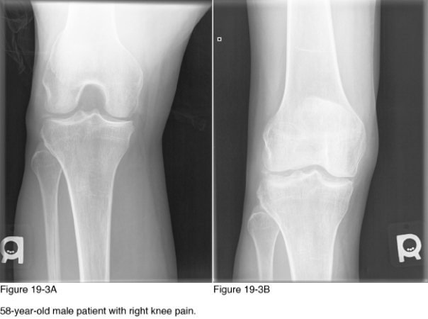
The underlying etiology of the disease observed in Figure 19-3A and Figure 19-3B is__________.
A) Autoimmune disease
B) Infection
C) Degenerative osteoarthritis
D) Endocrine/metabolic disease
Question
Which radiographic pattern of disease is representative in Figure 19-3A and Figure 19-3B?
A) Arthritides
B) Generalized osteosclerosis
C) Dwarfism
D) Polyostotic bone lesions
Question
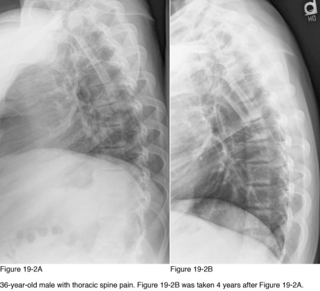
Which radiographic pattern of disease is observed in Figure 19-2?
A) Generalized osteosclerosis
B) Arthritides
C) Soft tissue calcification
D) Soft tissue ossification
Question
The salient radiographic abnormality and Figure 19-3A and Figure 19-3B is observed in the __________.
A) Femorotibial joint space
B) Femoral and tibial metaphyses
C) Musculature and subcutaneous tissues
D) Subchondral cortical and medullary bone
Question
For what other areas of the skeleton should radiographic examination be considered to assess full extent of this patient's disease?
A) Chest
B) Knees and forearms
C) Skull and sinuses
D) Cervical and thoracic spine
Question
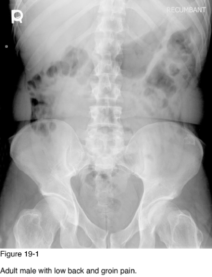
The radiographic abnormalities observed in Figure 19-1 are the result of __________.
A) Faulty enchondral ossification
B) Multiple osteochondromas
C) Metastasis
D) Heavy-metal exposure
Question
Chronic failure of which organ(s) is responsible for the changes observed between Figure 19-2A and Figure 19-2B?
A) Lymphatics
B) Lungs
C) Heart
D) Kidneys
Question
Development of pain in the regions of involvement with enlargement of the lesions is a sign of __________.
A) Metastasis
B) Pathologic fracture
C) Malignant degeneration
D) Accessory bursa formation
Question
Which laboratory value is likely to be elevated in this patient?
A) Alkaline phosphatase
B) Urinary hydroxyproline
C) Serum calcium
D) Acid phosphatase
Question
At what age are the lesions observed in Figure 19-1 generally diagnosed?
A) Younger than 20
B) 20-40
C) 40-60
D) Older than 60
Question
The primary site of abnormality in Figure 19-2B is observed within the __________.
A) Lung parenchyma
B) Ribs
C) Vertebral bodies adjacent to the endplates
D) Centrally within the vertebral bodies
Question
Which of the following general skeletal radiographic patterns of disease is evident in Figure 19-1?
A) Acetabular protrusion
B) Dwarfism
C) Generalized osteosclerosis
D) Polyostotic bone lesions
Question
Which of the following may be expected in radiographic examination of the hands of this patient?
A) Osteosclerotic bone lesions
B) Subperiosteal bone resorption
C) Changes in the size and shape of the epiphyses
D) Uniform joint space narrowing
Question
Which of the following diagnostic studies provide greatest sensitivity in assessing bone mineral density?
A) Bone scan
B) Digital radiography
C) SPECT
D) DEXA
Question
Which radiographic views are provided in Figure 19-3A and Figure 19-3B?
A) PA knee and tunnel
B) AP knee and tunnel
C) Merchant and AP
D) Sunrise and tunnel
Question
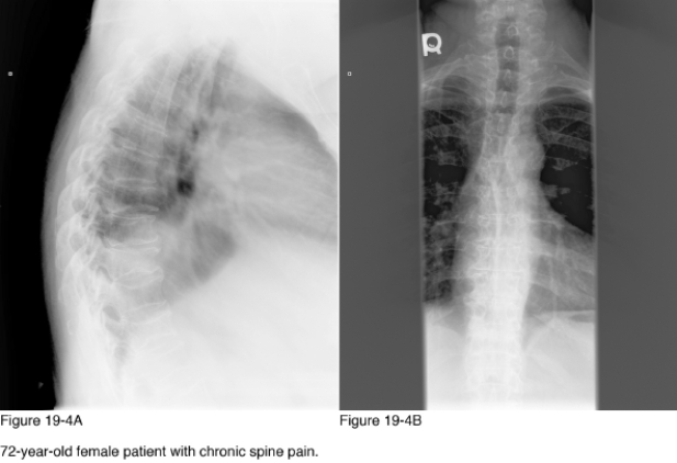
The endplate alterations observed in Figure 19-4A and Figure 19-4B are representative of __________.
A) Stress fractures
B) Insufficiency fractures
C) Congenital vertebral variant
D) Osteoarthritis
Question
The most common cost to the abnormalities observed in Figure 19-4A and Figure 19-4B is __________.
A) Autoimmune
B) Genetics
C) Age-related changes in bone composition
D) Nutritional disorder
Question
Which of the following treatments is not beneficial in addressing the underlying cause of the abnormalities observed in Figure 19-4A and Figure 19-4B?
A) Corticosteroids
B) Calcium supplementation
C) Calcitonin
D) Weight-bearing exercise
Question
Which of the following tests is considered diagnostic for this patient's disease?
A) Biopsy
B) Joint aspiration with microscopic assessment
C) Blood laboratory
D) Magnetic resonance imaging
Question
Which radiographic pattern of disease is observed in Figure 19-4A and Figure 19-4B?
A) Dwarfism
B) Generalized osteoporosis
C) Generalized osteosclerosis
D) Soft tissue calcification

Unlock Deck
Sign up to unlock the cards in this deck!
Unlock Deck
Unlock Deck
1/20
Play
Full screen (f)
Deck 19: General Skeletal Patterns
1
The underlying etiology of the disease observed in Figure 19-3A and Figure 19-3B is__________.
A) Autoimmune disease
B) Infection
C) Degenerative osteoarthritis
D) Endocrine/metabolic disease
Endocrine/metabolic disease
2
Which radiographic pattern of disease is representative in Figure 19-3A and Figure 19-3B?
A) Arthritides
B) Generalized osteosclerosis
C) Dwarfism
D) Polyostotic bone lesions
Arthritides
3
Which radiographic pattern of disease is observed in Figure 19-2?
A) Generalized osteosclerosis
B) Arthritides
C) Soft tissue calcification
D) Soft tissue ossification
Generalized osteosclerosis
4
The salient radiographic abnormality and Figure 19-3A and Figure 19-3B is observed in the __________.
A) Femorotibial joint space
B) Femoral and tibial metaphyses
C) Musculature and subcutaneous tissues
D) Subchondral cortical and medullary bone
Unlock Deck
Unlock for access to all 20 flashcards in this deck.
Unlock Deck
k this deck
5
For what other areas of the skeleton should radiographic examination be considered to assess full extent of this patient's disease?
A) Chest
B) Knees and forearms
C) Skull and sinuses
D) Cervical and thoracic spine
Unlock Deck
Unlock for access to all 20 flashcards in this deck.
Unlock Deck
k this deck
6
The radiographic abnormalities observed in Figure 19-1 are the result of __________.
A) Faulty enchondral ossification
B) Multiple osteochondromas
C) Metastasis
D) Heavy-metal exposure
Unlock Deck
Unlock for access to all 20 flashcards in this deck.
Unlock Deck
k this deck
7
Chronic failure of which organ(s) is responsible for the changes observed between Figure 19-2A and Figure 19-2B?
A) Lymphatics
B) Lungs
C) Heart
D) Kidneys
Unlock Deck
Unlock for access to all 20 flashcards in this deck.
Unlock Deck
k this deck
8
Development of pain in the regions of involvement with enlargement of the lesions is a sign of __________.
A) Metastasis
B) Pathologic fracture
C) Malignant degeneration
D) Accessory bursa formation
Unlock Deck
Unlock for access to all 20 flashcards in this deck.
Unlock Deck
k this deck
9
Which laboratory value is likely to be elevated in this patient?
A) Alkaline phosphatase
B) Urinary hydroxyproline
C) Serum calcium
D) Acid phosphatase
Unlock Deck
Unlock for access to all 20 flashcards in this deck.
Unlock Deck
k this deck
10
At what age are the lesions observed in Figure 19-1 generally diagnosed?
A) Younger than 20
B) 20-40
C) 40-60
D) Older than 60
Unlock Deck
Unlock for access to all 20 flashcards in this deck.
Unlock Deck
k this deck
11
The primary site of abnormality in Figure 19-2B is observed within the __________.
A) Lung parenchyma
B) Ribs
C) Vertebral bodies adjacent to the endplates
D) Centrally within the vertebral bodies
Unlock Deck
Unlock for access to all 20 flashcards in this deck.
Unlock Deck
k this deck
12
Which of the following general skeletal radiographic patterns of disease is evident in Figure 19-1?
A) Acetabular protrusion
B) Dwarfism
C) Generalized osteosclerosis
D) Polyostotic bone lesions
Unlock Deck
Unlock for access to all 20 flashcards in this deck.
Unlock Deck
k this deck
13
Which of the following may be expected in radiographic examination of the hands of this patient?
A) Osteosclerotic bone lesions
B) Subperiosteal bone resorption
C) Changes in the size and shape of the epiphyses
D) Uniform joint space narrowing
Unlock Deck
Unlock for access to all 20 flashcards in this deck.
Unlock Deck
k this deck
14
Which of the following diagnostic studies provide greatest sensitivity in assessing bone mineral density?
A) Bone scan
B) Digital radiography
C) SPECT
D) DEXA
Unlock Deck
Unlock for access to all 20 flashcards in this deck.
Unlock Deck
k this deck
15
Which radiographic views are provided in Figure 19-3A and Figure 19-3B?
A) PA knee and tunnel
B) AP knee and tunnel
C) Merchant and AP
D) Sunrise and tunnel
Unlock Deck
Unlock for access to all 20 flashcards in this deck.
Unlock Deck
k this deck
16
The endplate alterations observed in Figure 19-4A and Figure 19-4B are representative of __________.
A) Stress fractures
B) Insufficiency fractures
C) Congenital vertebral variant
D) Osteoarthritis
Unlock Deck
Unlock for access to all 20 flashcards in this deck.
Unlock Deck
k this deck
17
The most common cost to the abnormalities observed in Figure 19-4A and Figure 19-4B is __________.
A) Autoimmune
B) Genetics
C) Age-related changes in bone composition
D) Nutritional disorder
Unlock Deck
Unlock for access to all 20 flashcards in this deck.
Unlock Deck
k this deck
18
Which of the following treatments is not beneficial in addressing the underlying cause of the abnormalities observed in Figure 19-4A and Figure 19-4B?
A) Corticosteroids
B) Calcium supplementation
C) Calcitonin
D) Weight-bearing exercise
Unlock Deck
Unlock for access to all 20 flashcards in this deck.
Unlock Deck
k this deck
19
Which of the following tests is considered diagnostic for this patient's disease?
A) Biopsy
B) Joint aspiration with microscopic assessment
C) Blood laboratory
D) Magnetic resonance imaging
Unlock Deck
Unlock for access to all 20 flashcards in this deck.
Unlock Deck
k this deck
20
Which radiographic pattern of disease is observed in Figure 19-4A and Figure 19-4B?
A) Dwarfism
B) Generalized osteoporosis
C) Generalized osteosclerosis
D) Soft tissue calcification
Unlock Deck
Unlock for access to all 20 flashcards in this deck.
Unlock Deck
k this deck
Unlock Deck
Unlock for access to all 20 flashcards in this deck.


