Deck 17: Spine Patterns
Full screen (f)
Question
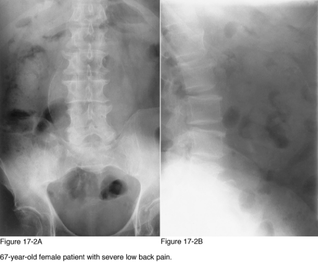
Which infection is most likely to result in paraspinal calcification secondary to abscess formation?
A) Salmonella
B) Staphylococcus aureus
C) Echinococcus granulosus
D) Mycobacterium tuberculosis
Question
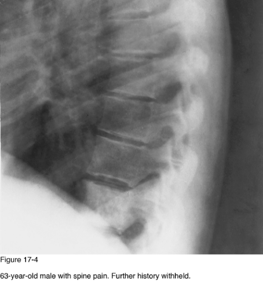
If Paget disease was the underlying cause for the abnormality present in Figure 17-4, which radiographic finding would likely also be observed?
A) Vertebra plana
B) Narrowed intervertebral disc
C) Vertebral enlargement
D) Paraspinal ligament ossification
Question
If the sacroiliac joint in this patient were destroyed, which of the following diagnoses should be considered?
A) Neuropathic spine
B) Trauma
C) Seronegative spondyloarthropathy
D) Diffuse idiopathic skeletal hyperostosis
Question

In patients with sickle cell disease, the underlying cause of vertebral body morphology similar to that observed in Figure 17-1 is __________?
A) Avascular necrosis
B) Insufficiency fracture
C) Underperfusion of the vertebral body
D) Prolapse of the nucleus pulposus
Question
Which of the following diagnostic imaging modalities would be the most helpful in identifying additional sites of involvement in the skeleton?
A) Diagnostic ultrasound
B) Computed tomography
C) Radionuclide scintigraphy
D) Skeletal x-ray survey
Question
Which of the following is the most common etiology for the change in vertebral body shape identified in Figure 17-1?
A) Osteoporosis
B) Thalassemia
C) Osteomalacia
D) Congenital abnormality
Question
Which of the following diagnoses would be most likely if radiographs showed multiple levels of endobones?
A) Albers-Schonberg disease
B) Paget disease
C) Fractures
D) Myelofibrosis
Question
In Figure 17-2A, the curvilinear calcifications are diagnostic for:
A) Paraspinal abscess
B) Aneurysm
C) Expansile spine lesion
D) Neurogenic tumor
Question

Patients older than __________ years of age are typically affected by the pathology observed in these radiographs.
A) 10
B) 20
C) 30
D) 40
Question
What pattern of vertebral body shape is present in Figure 17-1?
A) Wedged
B) Biconcave
C) Posterior scalloped
D) Pancake
Question
Which radiographic sign is present in Figure 17-3?
A) Winking owl
B) Fish mouth vertebra
C) Empty vertebra
D) Ivory vertebra
Question
Which of the following is the most important radiographic pattern of disease present in Figure 17-2?
A) Paraspinal mass
B) Narrow intervertebral disc
C) Sacroiliac joint disease
D) Vertebral endplate alteration
Question
Which of the following is the least likely cause of the abnormality in Figure 17-4?
A) Paget disease
B) Lymphoma
C) Osteoblastic metastasis
D) Osteopetrosis
Question
Of the following, which is the most common cause of the salient abnormality in Figure 17-3?
A) Chordoma
B) Hemangioma
C) Metastasis
D) Multiple myeloma
Question
Which of the following diseases is a genetic disorder resulting in defective collagen metabolism that may result in osteopenic vertebrae with this morphology?
A) Renal osteodystrophy
B) Homocystinuria
C) Gaucher disease
D) Notochordal persistency
Question
Which radiographic pattern of disease is represented in Figure 17-4?
A) Osteolytic spine
B) Radiodense ivory vertebra
C) Narrow intervertebral disc
D) Anterior vertebral body scalloping
Question
The bony outgrowths projecting from the endplates of the lumbar spine are representative of __________.
A) Ankylosing spondylitis
B) Diffuse idiopathic skeletal hyperostosis
C) Psoriatic arthropathy
D) Spondylosis deformans
Question
The most common reason for this vertebral body morphology in a 75-year-old female is __________.
A) Malnutrition
B) Senile osteoporosis
C) Osteomalacia
D) Hyperparathyroidism
Question
Which of the following is the most significant radiographic pattern of disease present in Figure 17-3?
A) Narrow intervertebral disc
B) Anterior scalloped vertebra
C) Paraspinal mass
D) Osteolytic lesion of the spine
Question
If an expansile lesion were observed in a 30-year-old patient in the same area of concern as in Figure 17-3, which of the following diagnoses would be the most likely diagnosis?
A) Aneurysmal bone cyst
B) Eosinophilic granuloma
C) Lymphoma
D) Leukemia

Unlock Deck
Sign up to unlock the cards in this deck!
Unlock Deck
Unlock Deck
1/20
Play
Full screen (f)
Deck 17: Spine Patterns
1
Which infection is most likely to result in paraspinal calcification secondary to abscess formation?
A) Salmonella
B) Staphylococcus aureus
C) Echinococcus granulosus
D) Mycobacterium tuberculosis
Mycobacterium tuberculosis
2
If Paget disease was the underlying cause for the abnormality present in Figure 17-4, which radiographic finding would likely also be observed?
A) Vertebra plana
B) Narrowed intervertebral disc
C) Vertebral enlargement
D) Paraspinal ligament ossification
Vertebral enlargement
3
If the sacroiliac joint in this patient were destroyed, which of the following diagnoses should be considered?
A) Neuropathic spine
B) Trauma
C) Seronegative spondyloarthropathy
D) Diffuse idiopathic skeletal hyperostosis
Seronegative spondyloarthropathy
4
In patients with sickle cell disease, the underlying cause of vertebral body morphology similar to that observed in Figure 17-1 is __________?
A) Avascular necrosis
B) Insufficiency fracture
C) Underperfusion of the vertebral body
D) Prolapse of the nucleus pulposus
Unlock Deck
Unlock for access to all 20 flashcards in this deck.
Unlock Deck
k this deck
5
Which of the following diagnostic imaging modalities would be the most helpful in identifying additional sites of involvement in the skeleton?
A) Diagnostic ultrasound
B) Computed tomography
C) Radionuclide scintigraphy
D) Skeletal x-ray survey
Unlock Deck
Unlock for access to all 20 flashcards in this deck.
Unlock Deck
k this deck
6
Which of the following is the most common etiology for the change in vertebral body shape identified in Figure 17-1?
A) Osteoporosis
B) Thalassemia
C) Osteomalacia
D) Congenital abnormality
Unlock Deck
Unlock for access to all 20 flashcards in this deck.
Unlock Deck
k this deck
7
Which of the following diagnoses would be most likely if radiographs showed multiple levels of endobones?
A) Albers-Schonberg disease
B) Paget disease
C) Fractures
D) Myelofibrosis
Unlock Deck
Unlock for access to all 20 flashcards in this deck.
Unlock Deck
k this deck
8
In Figure 17-2A, the curvilinear calcifications are diagnostic for:
A) Paraspinal abscess
B) Aneurysm
C) Expansile spine lesion
D) Neurogenic tumor
Unlock Deck
Unlock for access to all 20 flashcards in this deck.
Unlock Deck
k this deck
9
Patients older than __________ years of age are typically affected by the pathology observed in these radiographs.
A) 10
B) 20
C) 30
D) 40
Unlock Deck
Unlock for access to all 20 flashcards in this deck.
Unlock Deck
k this deck
10
What pattern of vertebral body shape is present in Figure 17-1?
A) Wedged
B) Biconcave
C) Posterior scalloped
D) Pancake
Unlock Deck
Unlock for access to all 20 flashcards in this deck.
Unlock Deck
k this deck
11
Which radiographic sign is present in Figure 17-3?
A) Winking owl
B) Fish mouth vertebra
C) Empty vertebra
D) Ivory vertebra
Unlock Deck
Unlock for access to all 20 flashcards in this deck.
Unlock Deck
k this deck
12
Which of the following is the most important radiographic pattern of disease present in Figure 17-2?
A) Paraspinal mass
B) Narrow intervertebral disc
C) Sacroiliac joint disease
D) Vertebral endplate alteration
Unlock Deck
Unlock for access to all 20 flashcards in this deck.
Unlock Deck
k this deck
13
Which of the following is the least likely cause of the abnormality in Figure 17-4?
A) Paget disease
B) Lymphoma
C) Osteoblastic metastasis
D) Osteopetrosis
Unlock Deck
Unlock for access to all 20 flashcards in this deck.
Unlock Deck
k this deck
14
Of the following, which is the most common cause of the salient abnormality in Figure 17-3?
A) Chordoma
B) Hemangioma
C) Metastasis
D) Multiple myeloma
Unlock Deck
Unlock for access to all 20 flashcards in this deck.
Unlock Deck
k this deck
15
Which of the following diseases is a genetic disorder resulting in defective collagen metabolism that may result in osteopenic vertebrae with this morphology?
A) Renal osteodystrophy
B) Homocystinuria
C) Gaucher disease
D) Notochordal persistency
Unlock Deck
Unlock for access to all 20 flashcards in this deck.
Unlock Deck
k this deck
16
Which radiographic pattern of disease is represented in Figure 17-4?
A) Osteolytic spine
B) Radiodense ivory vertebra
C) Narrow intervertebral disc
D) Anterior vertebral body scalloping
Unlock Deck
Unlock for access to all 20 flashcards in this deck.
Unlock Deck
k this deck
17
The bony outgrowths projecting from the endplates of the lumbar spine are representative of __________.
A) Ankylosing spondylitis
B) Diffuse idiopathic skeletal hyperostosis
C) Psoriatic arthropathy
D) Spondylosis deformans
Unlock Deck
Unlock for access to all 20 flashcards in this deck.
Unlock Deck
k this deck
18
The most common reason for this vertebral body morphology in a 75-year-old female is __________.
A) Malnutrition
B) Senile osteoporosis
C) Osteomalacia
D) Hyperparathyroidism
Unlock Deck
Unlock for access to all 20 flashcards in this deck.
Unlock Deck
k this deck
19
Which of the following is the most significant radiographic pattern of disease present in Figure 17-3?
A) Narrow intervertebral disc
B) Anterior scalloped vertebra
C) Paraspinal mass
D) Osteolytic lesion of the spine
Unlock Deck
Unlock for access to all 20 flashcards in this deck.
Unlock Deck
k this deck
20
If an expansile lesion were observed in a 30-year-old patient in the same area of concern as in Figure 17-3, which of the following diagnoses would be the most likely diagnosis?
A) Aneurysmal bone cyst
B) Eosinophilic granuloma
C) Lymphoma
D) Leukemia
Unlock Deck
Unlock for access to all 20 flashcards in this deck.
Unlock Deck
k this deck
Unlock Deck
Unlock for access to all 20 flashcards in this deck.


