Deck 18: Extremity Patterns
Full screen (f)
Question
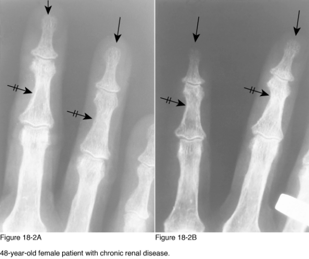
Which of the following radiographic diagnoses best accounts for the changes observed on the area marked by the crossed arrows in Figure 18-2?
A) Neurotrophic disease
B) Psoriatic arthritis
C) Scleroderma
D) Hyperparathyroidism
Question
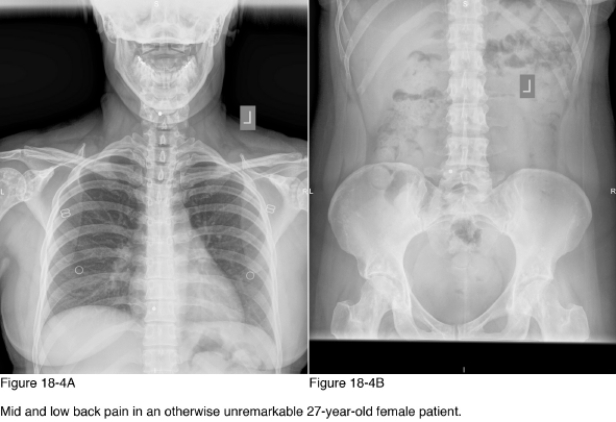
What further action/diagnostic testing is necessary to manage this patient specific to the radiographic abnormalities noted in Figure 18-4?
A) No further action
B) Radiographic survey of the major joints
C) Referral to an oncologist
D) Referral to a rheumatologist
Question
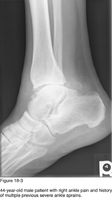
Which of the following radiographic findings supports your answer to the most likely cause of the abnormalities observed in Figure 18-3?
A) Nonuniform reduction of joint spacing
B) Polyarticular involvement
C) Soft tissue swelling
D) Joint disintegration
Question
Soft tissue calcifications in patients with the abnormality marked by the black arrows in Figure 18-2 are associated with __________ and __________.
A) Hyperparathyroidism, scleroderma
B) Sarcoidosis, thermal injury
C) Psoriatic arthritis, hyperparathyroidism
D) Scleroderma, arteriosclerosis obliterans
Question
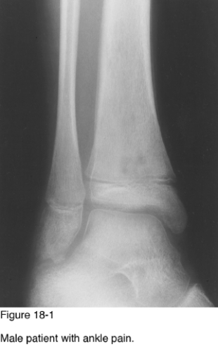
The location of the lesion and Figure 18-1 is best described as__________.
A) Metaphyseal cortex
B) Metaphyseal medullary cavity
C) Epiphyseal cortex
D) Intra-articular
Question
Which of the following diagnoses would be most likely if the radiographs demonstrate changes similar to those marked by the black arrows, with diffuse sclerosis observed in the bones of the hand in a patient with short stature?
A) Arteriosclerosis obliterans
B) Sarcoidosis
C) Pyknodysostosis
D) Progressive systemic sclerosis
Question
Which radiographic pattern of disease is noted in Figure 18-2, specific to the plain black arrows?
A) Acroosteolysis
B) Aggressive osteolytic lesions
C) Periosteal reaction
D) Change in size and shape of an epiphysis
Question
Which of the following is most likely to be the clinical complaint of the patient regarding his ankle?
A) No complaint
B) Polyarticular joint swelling
C) Acute pain with joint locking
D) Inability to bear weight
Question
With a history of mid and low back pain in an otherwise unremarkable 27-year-old female patient, which of the following is the best diagnosis of the abnormalities observed on the radiographs in Figure 18-4?
A) Foley areas disease
B) Osteonecrosis
C) Osteopoikilosis
D) Synovial chondrometaplasia
Question
Which radiographic pattern of disease is present Figure 18-1?
A) Change in size and shape of epiphysis
B) Cystic lesion of extremities and ribs
C) Radiolucent metaphyseal bands
D) Aggressive osteolytic lesions of the extremities and ribs
Question
Lesions observed in these radiographs are microscopically identical to a(n) __________.
A) Osteochondroma
B) Enostosis
C) Enchondroma
D) Nonossifying fibroma
Question
Which radiographic pattern of disease is observed in Figure 18-4?
A) Calcified intra-articular loose bodies
B) Aggressive osteolytic lesions of the ribs and extremities
C) Osteosclerotic bone lesions
D) Periosteal reactions
Question
Which radiographic finding in the spine is associated with the underlying disease observed in Figure 18-2?
A) Ivory vertebra
B) Rugger jersey
C) Sandwich vertebra
D) Vertebra plana
Question
Which of the following diagnoses accounts for this radiographic presentation?
A) Eosinophilic granuloma
B) Metastasis
C) Infection
D) Leukemia
Question
On the basis of Figure 18-1, what is the approximate age of this patient?
A) 0-5
B) 11-15
C) 21-25
D) 61-65
Question
The classic treatment for this lesion is __________.
A) Drainage, curettage, antibiotics
B) Amputation
C) Chemotherapy
D) This condition is self-resolving
Question
If similar radiographic presentation is observed in a patient older than 40 years of age with pain in the areas of radiographic abnormality, as well as personal history of previous malignancy, which of the following diagnostic studies would help assess for skeletal metastasis?
A) Diagnostic ultrasound
B) Skeletal x-ray survey
C) Fluoroscopy
D) Radionuclide scintigraphy
Question
Which of the following is the most likely cause of the abnormalities observed on these radiographs?
A) Neurotrophic joint disease
B) Crystal deposition disease
C) Infection
D) Degenerative joint disease
Question
A history of __________ is the most likely factor predisposing a patient to develop this presentation of joint disease.
A) Poor nutrition
B) Intravenous drug use
C) Trauma
D) Familial disease
Question
Which radiographic pattern of disease is observed in Figure 18-3?
A) Radiodense metaphyseal bands
B) Osteosclerotic bone lesions
C) Cystic lesions of the extremities and ribs
D) Calcified intra-articular loose bodies

Unlock Deck
Sign up to unlock the cards in this deck!
Unlock Deck
Unlock Deck
1/20
Play
Full screen (f)
Deck 18: Extremity Patterns
1
Which of the following radiographic diagnoses best accounts for the changes observed on the area marked by the crossed arrows in Figure 18-2?
A) Neurotrophic disease
B) Psoriatic arthritis
C) Scleroderma
D) Hyperparathyroidism
Hyperparathyroidism
2
What further action/diagnostic testing is necessary to manage this patient specific to the radiographic abnormalities noted in Figure 18-4?
A) No further action
B) Radiographic survey of the major joints
C) Referral to an oncologist
D) Referral to a rheumatologist
No further action
3
Which of the following radiographic findings supports your answer to the most likely cause of the abnormalities observed in Figure 18-3?
A) Nonuniform reduction of joint spacing
B) Polyarticular involvement
C) Soft tissue swelling
D) Joint disintegration
Nonuniform reduction of joint spacing
4
Soft tissue calcifications in patients with the abnormality marked by the black arrows in Figure 18-2 are associated with __________ and __________.
A) Hyperparathyroidism, scleroderma
B) Sarcoidosis, thermal injury
C) Psoriatic arthritis, hyperparathyroidism
D) Scleroderma, arteriosclerosis obliterans
Unlock Deck
Unlock for access to all 20 flashcards in this deck.
Unlock Deck
k this deck
5
The location of the lesion and Figure 18-1 is best described as__________.
A) Metaphyseal cortex
B) Metaphyseal medullary cavity
C) Epiphyseal cortex
D) Intra-articular
Unlock Deck
Unlock for access to all 20 flashcards in this deck.
Unlock Deck
k this deck
6
Which of the following diagnoses would be most likely if the radiographs demonstrate changes similar to those marked by the black arrows, with diffuse sclerosis observed in the bones of the hand in a patient with short stature?
A) Arteriosclerosis obliterans
B) Sarcoidosis
C) Pyknodysostosis
D) Progressive systemic sclerosis
Unlock Deck
Unlock for access to all 20 flashcards in this deck.
Unlock Deck
k this deck
7
Which radiographic pattern of disease is noted in Figure 18-2, specific to the plain black arrows?
A) Acroosteolysis
B) Aggressive osteolytic lesions
C) Periosteal reaction
D) Change in size and shape of an epiphysis
Unlock Deck
Unlock for access to all 20 flashcards in this deck.
Unlock Deck
k this deck
8
Which of the following is most likely to be the clinical complaint of the patient regarding his ankle?
A) No complaint
B) Polyarticular joint swelling
C) Acute pain with joint locking
D) Inability to bear weight
Unlock Deck
Unlock for access to all 20 flashcards in this deck.
Unlock Deck
k this deck
9
With a history of mid and low back pain in an otherwise unremarkable 27-year-old female patient, which of the following is the best diagnosis of the abnormalities observed on the radiographs in Figure 18-4?
A) Foley areas disease
B) Osteonecrosis
C) Osteopoikilosis
D) Synovial chondrometaplasia
Unlock Deck
Unlock for access to all 20 flashcards in this deck.
Unlock Deck
k this deck
10
Which radiographic pattern of disease is present Figure 18-1?
A) Change in size and shape of epiphysis
B) Cystic lesion of extremities and ribs
C) Radiolucent metaphyseal bands
D) Aggressive osteolytic lesions of the extremities and ribs
Unlock Deck
Unlock for access to all 20 flashcards in this deck.
Unlock Deck
k this deck
11
Lesions observed in these radiographs are microscopically identical to a(n) __________.
A) Osteochondroma
B) Enostosis
C) Enchondroma
D) Nonossifying fibroma
Unlock Deck
Unlock for access to all 20 flashcards in this deck.
Unlock Deck
k this deck
12
Which radiographic pattern of disease is observed in Figure 18-4?
A) Calcified intra-articular loose bodies
B) Aggressive osteolytic lesions of the ribs and extremities
C) Osteosclerotic bone lesions
D) Periosteal reactions
Unlock Deck
Unlock for access to all 20 flashcards in this deck.
Unlock Deck
k this deck
13
Which radiographic finding in the spine is associated with the underlying disease observed in Figure 18-2?
A) Ivory vertebra
B) Rugger jersey
C) Sandwich vertebra
D) Vertebra plana
Unlock Deck
Unlock for access to all 20 flashcards in this deck.
Unlock Deck
k this deck
14
Which of the following diagnoses accounts for this radiographic presentation?
A) Eosinophilic granuloma
B) Metastasis
C) Infection
D) Leukemia
Unlock Deck
Unlock for access to all 20 flashcards in this deck.
Unlock Deck
k this deck
15
On the basis of Figure 18-1, what is the approximate age of this patient?
A) 0-5
B) 11-15
C) 21-25
D) 61-65
Unlock Deck
Unlock for access to all 20 flashcards in this deck.
Unlock Deck
k this deck
16
The classic treatment for this lesion is __________.
A) Drainage, curettage, antibiotics
B) Amputation
C) Chemotherapy
D) This condition is self-resolving
Unlock Deck
Unlock for access to all 20 flashcards in this deck.
Unlock Deck
k this deck
17
If similar radiographic presentation is observed in a patient older than 40 years of age with pain in the areas of radiographic abnormality, as well as personal history of previous malignancy, which of the following diagnostic studies would help assess for skeletal metastasis?
A) Diagnostic ultrasound
B) Skeletal x-ray survey
C) Fluoroscopy
D) Radionuclide scintigraphy
Unlock Deck
Unlock for access to all 20 flashcards in this deck.
Unlock Deck
k this deck
18
Which of the following is the most likely cause of the abnormalities observed on these radiographs?
A) Neurotrophic joint disease
B) Crystal deposition disease
C) Infection
D) Degenerative joint disease
Unlock Deck
Unlock for access to all 20 flashcards in this deck.
Unlock Deck
k this deck
19
A history of __________ is the most likely factor predisposing a patient to develop this presentation of joint disease.
A) Poor nutrition
B) Intravenous drug use
C) Trauma
D) Familial disease
Unlock Deck
Unlock for access to all 20 flashcards in this deck.
Unlock Deck
k this deck
20
Which radiographic pattern of disease is observed in Figure 18-3?
A) Radiodense metaphyseal bands
B) Osteosclerotic bone lesions
C) Cystic lesions of the extremities and ribs
D) Calcified intra-articular loose bodies
Unlock Deck
Unlock for access to all 20 flashcards in this deck.
Unlock Deck
k this deck
Unlock Deck
Unlock for access to all 20 flashcards in this deck.


