Deck 16: Skull Patterns
Full screen (f)
Question
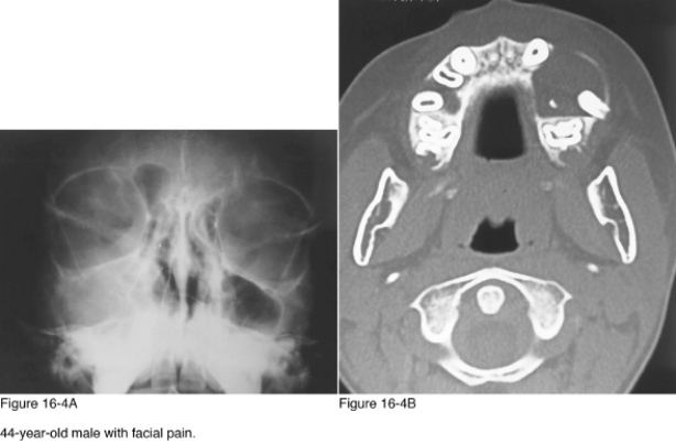
In Figure 16-4A there is opacification of the __________ sinus.
A) Frontal
B) Ethmoid
C) Sphenoid
D) Maxillary
Question
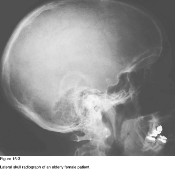
Chronic disease of the __________ may produce the appearance of the skull in Figure 16-3.
A) Lungs
B) Prostate
C) Kidneys
D) Adrenals
Question
Which of the following best describes the diagnostic imaging study represented by Figure 16-4B?
A) Axial computed tomography through the maxilla
B) Axial computed tomography through the mandible
C) Axial magnetic resonance imaging through the maxilla
D) Axial magnetic resonance imaging through the mandible
Question
Which of the following diagnoses best accounts for the well-circumscribed cyst containing the crown of an unerupted tooth in Figure 16-4B?
A) Fibrous dysplasia
B) Dentigerous cyst
C) Infection
D) Solitary bone cyst
Question
The granular appearance of the skull in Figure 16-3 is best described as __________.
A) Salt and pepper
B) Punched out
C) Hair on end
D) Osteoporosis circumscripta
Question
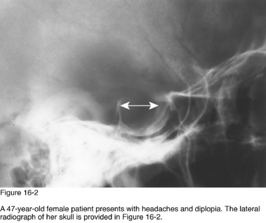
Enlargement of the sella turcica secondary to entrasellar extension of the subarachnoid space through defect in the diaphragm sellae describes __________.
A) Increased intracranial pressure
B) Aneurysm of the carotid vessel
C) Meningioma
D) Empty sella syndrome
Question
Which of the following radiographic patterns of disease is represented in Figure 16-3?
A) Enlarged sella turcica
B) Basilar impression
C) Diffuse demineralization
D) Cystic lesion of the mandible
Question
What type of pituitary adenoma is likely to result in enlargement of the sella turcica in patients with acromegaly?
A) Eosinophilic
B) Chromophobe
C) Basophilic
D) Craniopharyngioma
Question
The anteroposterior dimension of the sella turcica should not exceed __________mm.
A) 8
B) 12
C) 16
D) 18
Question
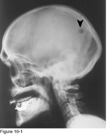
Which of the following radiographic patterns of disease is represented in Figure 16-1?
A) Basilar invagination
B) Intracranial calcifications
C) Radiodense mandible lesions
D) Osteolytic defect of the skull
Question
Which of the following radiographic patterns of disease is represented in Figure 16-4A?
A) Mass in the paranasal sinus
B) Radiodense mandible lesions
C) Intracranial calcifications
D) Cystic lesions of the mandible and skull
Question
Sickle-cell anemia and thalassemia may produce findings observed in this radiograph due to __________.
A) Metastatic disease
B) Bone infarcts
C) Endocrinopathy
D) Marrow hyperplasia
Question
Of the following, which is the most common disease producing the radiographic appearance of Figure 16-4A?
A) Polypoid rhinosinusitis
B) Acute sinusitis
C) Fibrous dysplasia
D) Wegener granulomatosus
Question
Aneurysm of which of the following arteries may produce enlargement of the sella turcica?
A) External carotid
B) Internal carotid
C) Middle meningeal
D) Basilar
Question
Of the following, which is the most common cause of the lesion observed in Figure 16-1?
A) Metastasis
B) Necrosis
C) Encephalocele
D) Primary malignancy
Question
Which of the following diagnostic imaging studies would be most appropriate if the patient also had a history of primary breast malignancy with mastectomy 2 years before this examination?
A) Skeletal x-ray survey
B) CT examination of the head
C) MRI examination of the head
D) Radionuclide scintigraphy
Question
Which of the following diagnoses would best explain the lesion, if on biopsy it was determined to contain ectodermal and mesodermal tissue?
A) Histiocytosis X
B) Multiple myeloma
C) Dermoid
D) Leptomeningeal cyst
Question
Of the following, which is the most likely reason for enlargement of the sella turcica in this patient?
A) Increased intracranial pressure
B) Meningioma
C) Chordoma
D) Pituitary tumor
Question
If the lesion in Figure 16-1 was surrounded by an osteosclerotic border, it would be known as a __________ ?
A) Button sequestration
B) Punched-out lesion
C) Donut lesion
D) Raindrop lesion
Question
Of the following, the most common cause of this presentation is ___________.
A) Metastatic bone disease
B) Osteoporosis
C) Multiple myeloma
D) Paget disease

Unlock Deck
Sign up to unlock the cards in this deck!
Unlock Deck
Unlock Deck
1/20
Play
Full screen (f)
Deck 16: Skull Patterns
1
In Figure 16-4A there is opacification of the __________ sinus.
A) Frontal
B) Ethmoid
C) Sphenoid
D) Maxillary
Maxillary
2
Chronic disease of the __________ may produce the appearance of the skull in Figure 16-3.
A) Lungs
B) Prostate
C) Kidneys
D) Adrenals
Kidneys
3
Which of the following best describes the diagnostic imaging study represented by Figure 16-4B?
A) Axial computed tomography through the maxilla
B) Axial computed tomography through the mandible
C) Axial magnetic resonance imaging through the maxilla
D) Axial magnetic resonance imaging through the mandible
Axial computed tomography through the maxilla
4
Which of the following diagnoses best accounts for the well-circumscribed cyst containing the crown of an unerupted tooth in Figure 16-4B?
A) Fibrous dysplasia
B) Dentigerous cyst
C) Infection
D) Solitary bone cyst
Unlock Deck
Unlock for access to all 20 flashcards in this deck.
Unlock Deck
k this deck
5
The granular appearance of the skull in Figure 16-3 is best described as __________.
A) Salt and pepper
B) Punched out
C) Hair on end
D) Osteoporosis circumscripta
Unlock Deck
Unlock for access to all 20 flashcards in this deck.
Unlock Deck
k this deck
6
Enlargement of the sella turcica secondary to entrasellar extension of the subarachnoid space through defect in the diaphragm sellae describes __________.
A) Increased intracranial pressure
B) Aneurysm of the carotid vessel
C) Meningioma
D) Empty sella syndrome
Unlock Deck
Unlock for access to all 20 flashcards in this deck.
Unlock Deck
k this deck
7
Which of the following radiographic patterns of disease is represented in Figure 16-3?
A) Enlarged sella turcica
B) Basilar impression
C) Diffuse demineralization
D) Cystic lesion of the mandible
Unlock Deck
Unlock for access to all 20 flashcards in this deck.
Unlock Deck
k this deck
8
What type of pituitary adenoma is likely to result in enlargement of the sella turcica in patients with acromegaly?
A) Eosinophilic
B) Chromophobe
C) Basophilic
D) Craniopharyngioma
Unlock Deck
Unlock for access to all 20 flashcards in this deck.
Unlock Deck
k this deck
9
The anteroposterior dimension of the sella turcica should not exceed __________mm.
A) 8
B) 12
C) 16
D) 18
Unlock Deck
Unlock for access to all 20 flashcards in this deck.
Unlock Deck
k this deck
10
Which of the following radiographic patterns of disease is represented in Figure 16-1?
A) Basilar invagination
B) Intracranial calcifications
C) Radiodense mandible lesions
D) Osteolytic defect of the skull
Unlock Deck
Unlock for access to all 20 flashcards in this deck.
Unlock Deck
k this deck
11
Which of the following radiographic patterns of disease is represented in Figure 16-4A?
A) Mass in the paranasal sinus
B) Radiodense mandible lesions
C) Intracranial calcifications
D) Cystic lesions of the mandible and skull
Unlock Deck
Unlock for access to all 20 flashcards in this deck.
Unlock Deck
k this deck
12
Sickle-cell anemia and thalassemia may produce findings observed in this radiograph due to __________.
A) Metastatic disease
B) Bone infarcts
C) Endocrinopathy
D) Marrow hyperplasia
Unlock Deck
Unlock for access to all 20 flashcards in this deck.
Unlock Deck
k this deck
13
Of the following, which is the most common disease producing the radiographic appearance of Figure 16-4A?
A) Polypoid rhinosinusitis
B) Acute sinusitis
C) Fibrous dysplasia
D) Wegener granulomatosus
Unlock Deck
Unlock for access to all 20 flashcards in this deck.
Unlock Deck
k this deck
14
Aneurysm of which of the following arteries may produce enlargement of the sella turcica?
A) External carotid
B) Internal carotid
C) Middle meningeal
D) Basilar
Unlock Deck
Unlock for access to all 20 flashcards in this deck.
Unlock Deck
k this deck
15
Of the following, which is the most common cause of the lesion observed in Figure 16-1?
A) Metastasis
B) Necrosis
C) Encephalocele
D) Primary malignancy
Unlock Deck
Unlock for access to all 20 flashcards in this deck.
Unlock Deck
k this deck
16
Which of the following diagnostic imaging studies would be most appropriate if the patient also had a history of primary breast malignancy with mastectomy 2 years before this examination?
A) Skeletal x-ray survey
B) CT examination of the head
C) MRI examination of the head
D) Radionuclide scintigraphy
Unlock Deck
Unlock for access to all 20 flashcards in this deck.
Unlock Deck
k this deck
17
Which of the following diagnoses would best explain the lesion, if on biopsy it was determined to contain ectodermal and mesodermal tissue?
A) Histiocytosis X
B) Multiple myeloma
C) Dermoid
D) Leptomeningeal cyst
Unlock Deck
Unlock for access to all 20 flashcards in this deck.
Unlock Deck
k this deck
18
Of the following, which is the most likely reason for enlargement of the sella turcica in this patient?
A) Increased intracranial pressure
B) Meningioma
C) Chordoma
D) Pituitary tumor
Unlock Deck
Unlock for access to all 20 flashcards in this deck.
Unlock Deck
k this deck
19
If the lesion in Figure 16-1 was surrounded by an osteosclerotic border, it would be known as a __________ ?
A) Button sequestration
B) Punched-out lesion
C) Donut lesion
D) Raindrop lesion
Unlock Deck
Unlock for access to all 20 flashcards in this deck.
Unlock Deck
k this deck
20
Of the following, the most common cause of this presentation is ___________.
A) Metastatic bone disease
B) Osteoporosis
C) Multiple myeloma
D) Paget disease
Unlock Deck
Unlock for access to all 20 flashcards in this deck.
Unlock Deck
k this deck
Unlock Deck
Unlock for access to all 20 flashcards in this deck.


