Deck 4: Vision
Full screen (f)
Question
Question
Question
Question
Question
Question
Question
Figure Q4-14 shows the currents from a rod and a cone in response to increasing magnitude of light flashes. How do these responses contribute to the higher sensitivity in low light and higher acuity in daylight conditions? 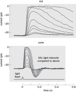 Figure Q4-14
Figure Q4-14 Question
Question
Question
Question
Question
Question
Question
Question
Question
Figure Q4-15 the spectral sensitivity of human cones. 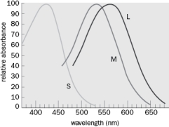 Figure Q4-15
A. What three wavelengths do we respond best to?
B. Which cones would respond to a very bright light whose wavelength was between 450 and 500 nm?
Figure Q4-15A. What three wavelengths do we respond best to?
B. Which cones would respond to a very bright light whose wavelength was between 450 and 500 nm?
Question
Question
Question
Our visual system has a remarkable dynamic range. When you go to a matinee movie (during the daytime) and walk out of the theater it is difficult to see. After a few minutes your eyes adapt to the new light levels. Some of the molecular mechanisms of light adaptation are known and most involve changes in Ca2+ concentration. 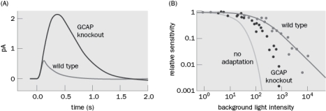 Figure Q4-13
A. Based on the data from the experiments shown in Figure Q4-13, what is one mechanism of adaptation? Figure Q4-13A shows the dark-adapted response to a flash of light in wild type and GCAP knockout mice. Figure Q4-13B shows the rod response to a flash of light relative to the same flash after dark adaptation.
B. What is the probable molecular mechanism of reduced light adaptation involving GCAP?
Figure Q4-13A. Based on the data from the experiments shown in Figure Q4-13, what is one mechanism of adaptation? Figure Q4-13A shows the dark-adapted response to a flash of light in wild type and GCAP knockout mice. Figure Q4-13B shows the rod response to a flash of light relative to the same flash after dark adaptation.
B. What is the probable molecular mechanism of reduced light adaptation involving GCAP?
Question
The responses of a single rod to light flashes were recorded in Figure Q4-7. 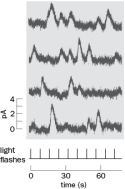 Figure Q4-7
A. What happened to the rod when it was stimulated with flashes of light?
B. Why is the lack of responses so important?
Figure Q4-7A. What happened to the rod when it was stimulated with flashes of light?
B. Why is the lack of responses so important?
Question
Question
Question
Question
An object of interest is to your right. The light from this object excites the temporal left eye and the nasal right eye as in Figure 4-32. 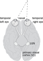 Figure 4-32
A. What happens to the organization of that image from the retina to the LGN and then to primary visual cortex? That is, what is the anatomical organization of the visual circuitry? Include the organization of the terminal projections in the LGN and cortex.
B. What would the projection pattern look like if you traced the path from two neighboring RGC from the left eye to the LGN and cortex?
Figure 4-32A. What happens to the organization of that image from the retina to the LGN and then to primary visual cortex? That is, what is the anatomical organization of the visual circuitry? Include the organization of the terminal projections in the LGN and cortex.
B. What would the projection pattern look like if you traced the path from two neighboring RGC from the left eye to the LGN and cortex?
Question
Question
Question
Question
Question
In the photoreceptor, stimulation of light results in an increase or decrease in glutamate release? Explain your answer.
Questions 4-25 and 4-26 refer to Figure Q4-25. You record from an on-center ganglion cell in complete darkness, present a light stimulus (horizontal bar), and elicit an increase in the number of action potentials in the RGC as in Figure Q4-25.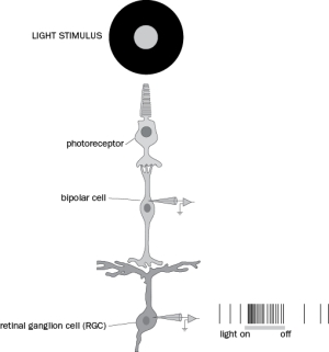 Figure Q4-25
Questions 4-25 and 4-26 refer to Figure Q4-25. You record from an on-center ganglion cell in complete darkness, present a light stimulus (horizontal bar), and elicit an increase in the number of action potentials in the RGC as in Figure Q4-25.
Figure Q4-25 Question
Question
Question
Question
Question
Question
Question
Question
Question
Question
Question
In Figure Q4-46, the middle temporal visual area (MT) was stimulated and the monkey indicated the direction of movement of dots in either the preferred or null direction. Below are the effects of MT stimulation from one animal. What was the effect of stimulation and how is that depicted on the graph? 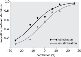 Figure Q4-46
Figure Q4-46 Question
Question
Question
Question
Question
Question
Question
Match between columns

Unlock Deck
Sign up to unlock the cards in this deck!
Unlock Deck
Unlock Deck
1/47
Play
Full screen (f)
Deck 4: Vision
1
In order to return to the dark state, a photoreceptor must undergo recovery. Recovery involves many steps. How does each step of phototransduction return to the dark state?
A. cGMP increase
B. Inactivation of transducin
C. Deactivation of rhodopsin
D. Retinal configuration
A. cGMP increase
B. Inactivation of transducin
C. Deactivation of rhodopsin
D. Retinal configuration
Light causes cGMP levels to decrease, which closes the CNG channels. When these channels close there is a decrease in the intracellular concentration of Ca2+. Guanylate cyclase activating protein (GCAP) activates guanylate cyclase in the absence of Ca2+, which increases cGMP levels.
B. Transducin is active when it is bound by Tα-GTP. Tα-GTP has intrinsic GTPase activity, which converts the alpha subunit to the GDP bound state and therefore no longer binds to transducin, which inactivates the protein. The conversion of GTP to GDP is facilitated by RGS9 (a GTPase activating protein), which activates the GTPase.
C. Rhodopsin is deactivated by rhodopsin kinase, which specifically binds to the phosphorylated rhodopsin protein. In addition arrestin competes for the Tα binding site on phosphorylated rhodopsin and prevents rhodopsin from activating transducin.
D. All-trans retinal must be converted back to 11-cis retinal. This happens in the pigment cells in the pigment epithelium.
2
When people get older the lens becomes stiffer and is not able to change shape as readily as when people are younger. Why would the inability of changing the angle of light through the lens cause objects (particularly those that are close) to become out of focus?
The lens helps focus light on the back of the retina. If the lens cannot change shape, light will not be focused on the back of the retina and the image will be out of focus. Reading glasses help change the focal length to refocus the image on the retina.
3
In photoreceptors, in the presence of light, injection of cGMP would:
(a) depolarize the cell.
(b) hyperpolarize the cell.
(c) not change the voltage across the membrane.
(a) depolarize the cell.
(b) hyperpolarize the cell.
(c) not change the voltage across the membrane.
(A)
Injection of cGMP would depolarize the cell. cGMP binds to the cyclic-nucleotide gated (CNG) channel and causes the channel to open. The reversal potential of the channel is about 0 mV so injection of cGMP would drive the voltage across the membrane toward 0mV, or depolarized.
Injection of cGMP would depolarize the cell. cGMP binds to the cyclic-nucleotide gated (CNG) channel and causes the channel to open. The reversal potential of the channel is about 0 mV so injection of cGMP would drive the voltage across the membrane toward 0mV, or depolarized.
4
Why are most laser pointers colored green or red, but not blue?
Unlock Deck
Unlock for access to all 47 flashcards in this deck.
Unlock Deck
k this deck
5
You are out in the middle of the desert star-gazing trying to see the North Star. What is the best way to look at the star? Why?
(a) With the fovea for better acuity
(b) From the side of your visual field for better sensitivity
(c) With the fovea for better contrast
(d) From the side of your visual field for better acuity
(a) With the fovea for better acuity
(b) From the side of your visual field for better sensitivity
(c) With the fovea for better contrast
(d) From the side of your visual field for better acuity
Unlock Deck
Unlock for access to all 47 flashcards in this deck.
Unlock Deck
k this deck
6
The M and L opsins are most similar in their amino acid structure. What does that suggest about their evolution?
Unlock Deck
Unlock for access to all 47 flashcards in this deck.
Unlock Deck
k this deck
7
Figure Q4-14 shows the currents from a rod and a cone in response to increasing magnitude of light flashes. How do these responses contribute to the higher sensitivity in low light and higher acuity in daylight conditions? Figure Q4-14
Figure Q4-14 Unlock Deck
Unlock for access to all 47 flashcards in this deck.
Unlock Deck
k this deck
8
What is the 'blind spot'? Select all that are correct.
(a) The place in the retina in which all axons exit the retina to go to the brain.
(b) The place the lens focuses light on the retina.
(c) The location in the retina where light has a direct path to the photoreceptors.
(d) The location in the retina with the highest density of capillaries.
(a) The place in the retina in which all axons exit the retina to go to the brain.
(b) The place the lens focuses light on the retina.
(c) The location in the retina where light has a direct path to the photoreceptors.
(d) The location in the retina with the highest density of capillaries.
Unlock Deck
Unlock for access to all 47 flashcards in this deck.
Unlock Deck
k this deck
9
What is the advantage of the retina using graded potentials? Explain.
Unlock Deck
Unlock for access to all 47 flashcards in this deck.
Unlock Deck
k this deck
10
Macular degeneration is damage to the macular, or center, of the retina. Why is loss of this area so devastating to vision?
Unlock Deck
Unlock for access to all 47 flashcards in this deck.
Unlock Deck
k this deck
11
What would happen if you blocked phosphodiesterase (PDE) and then stimulated the photoreceptor with light?
(a) The cell would depolarize.
(b) The cell would hyperpolarize.
(c) There would be no change in the voltage across the membrane.
(a) The cell would depolarize.
(b) The cell would hyperpolarize.
(c) There would be no change in the voltage across the membrane.
Unlock Deck
Unlock for access to all 47 flashcards in this deck.
Unlock Deck
k this deck
12
Fill in the missing words in the following paragraph.
Light is absorbed by the protein ______, which changes from _____ to a _____ configuration. This causes a structural change in the ____ protein. This conformational change allows the G protein called __________ to bind, and results in the exchange of _____ for GDP. The _______subunit of the G protein then activates the protein ______, which breaks down cGMP. The reduction in cGMP causes CNG channels to ______ and the cell to ______.
Light is absorbed by the protein ______, which changes from _____ to a _____ configuration. This causes a structural change in the ____ protein. This conformational change allows the G protein called __________ to bind, and results in the exchange of _____ for GDP. The _______subunit of the G protein then activates the protein ______, which breaks down cGMP. The reduction in cGMP causes CNG channels to ______ and the cell to ______.
Unlock Deck
Unlock for access to all 47 flashcards in this deck.
Unlock Deck
k this deck
13
Rhodopsin is what kind of protein?
(a) Voltage-dependent ion channel
(b) Ion co-transporter
(c) Na+ pump
(d) G-protein-coupled receptor
(a) Voltage-dependent ion channel
(b) Ion co-transporter
(c) Na+ pump
(d) G-protein-coupled receptor
Unlock Deck
Unlock for access to all 47 flashcards in this deck.
Unlock Deck
k this deck
14
Which cell types in the retina produce action potentials? Choose all that are correct.
(a) Photoreceptors
(b) Horizontal cells
(c) Bipolar cells
(d) Amacrine cells
(e) Retinal ganglion cells
(a) Photoreceptors
(b) Horizontal cells
(c) Bipolar cells
(d) Amacrine cells
(e) Retinal ganglion cells
Unlock Deck
Unlock for access to all 47 flashcards in this deck.
Unlock Deck
k this deck
15
Why are men more likely to be red/green color blind?
(a) Men have fewer photoreceptors with M and L opsins.
(b) M and L opsins are located close to each other on the X chromosome.
(c) Testosterone modulates the absorption of M and L opsins.
(d) M and L opsins probably evolved from a common ancestor.
How Are Signals from Rods and Cones Analyzed in the Retina?
Unlock Deck
Unlock for access to all 47 flashcards in this deck.
Unlock Deck
k this deck
16
Figure Q4-15 the spectral sensitivity of human cones. Figure Q4-15
A. What three wavelengths do we respond best to?
B. Which cones would respond to a very bright light whose wavelength was between 450 and 500 nm?
Figure Q4-15A. What three wavelengths do we respond best to?
B. Which cones would respond to a very bright light whose wavelength was between 450 and 500 nm?
Unlock Deck
Unlock for access to all 47 flashcards in this deck.
Unlock Deck
k this deck
17
What is the smallest number of photons of light that are able to elicit a change in membrane potential in a photoreceptor?
(a) 1
(b) 2
(c) 5
(d) 50
(a) 1
(b) 2
(c) 5
(d) 50
Unlock Deck
Unlock for access to all 47 flashcards in this deck.
Unlock Deck
k this deck
18
What is the fovea? Select all that are correct.
(a) The place in the retina in which all axons exit the retina to go to the brain.
(b) The place on the retina with the highest concentration of cones.
(c) The location in the retina where light has a direct path to the photoreceptors.
(d) The location in the retina with the highest density of capillaries.
Unlock Deck
Unlock for access to all 47 flashcards in this deck.
Unlock Deck
k this deck
19
Our visual system has a remarkable dynamic range. When you go to a matinee movie (during the daytime) and walk out of the theater it is difficult to see. After a few minutes your eyes adapt to the new light levels. Some of the molecular mechanisms of light adaptation are known and most involve changes in Ca2+ concentration. Figure Q4-13
A. Based on the data from the experiments shown in Figure Q4-13, what is one mechanism of adaptation? Figure Q4-13A shows the dark-adapted response to a flash of light in wild type and GCAP knockout mice. Figure Q4-13B shows the rod response to a flash of light relative to the same flash after dark adaptation.
B. What is the probable molecular mechanism of reduced light adaptation involving GCAP?
Figure Q4-13A. Based on the data from the experiments shown in Figure Q4-13, what is one mechanism of adaptation? Figure Q4-13A shows the dark-adapted response to a flash of light in wild type and GCAP knockout mice. Figure Q4-13B shows the rod response to a flash of light relative to the same flash after dark adaptation.
B. What is the probable molecular mechanism of reduced light adaptation involving GCAP?
Unlock Deck
Unlock for access to all 47 flashcards in this deck.
Unlock Deck
k this deck
20
The responses of a single rod to light flashes were recorded in Figure Q4-7. Figure Q4-7
A. What happened to the rod when it was stimulated with flashes of light?
B. Why is the lack of responses so important?
Figure Q4-7A. What happened to the rod when it was stimulated with flashes of light?
B. Why is the lack of responses so important?
Unlock Deck
Unlock for access to all 47 flashcards in this deck.
Unlock Deck
k this deck
21
How does the influence of horizontal cells help explain the optical illusion in the Mach bands in which the edge of a gray band appears darker on one side and lighter on the other?
Unlock Deck
Unlock for access to all 47 flashcards in this deck.
Unlock Deck
k this deck
22
Where does binocular input first occur in the visual system and why does it emerge at this location?
Unlock Deck
Unlock for access to all 47 flashcards in this deck.
Unlock Deck
k this deck
23
Besides rods and cones, what is the third type of photoreceptor in the retina?
Unlock Deck
Unlock for access to all 47 flashcards in this deck.
Unlock Deck
k this deck
24
An object of interest is to your right. The light from this object excites the temporal left eye and the nasal right eye as in Figure 4-32. Figure 4-32
A. What happens to the organization of that image from the retina to the LGN and then to primary visual cortex? That is, what is the anatomical organization of the visual circuitry? Include the organization of the terminal projections in the LGN and cortex.
B. What would the projection pattern look like if you traced the path from two neighboring RGC from the left eye to the LGN and cortex?
Figure 4-32A. What happens to the organization of that image from the retina to the LGN and then to primary visual cortex? That is, what is the anatomical organization of the visual circuitry? Include the organization of the terminal projections in the LGN and cortex.
B. What would the projection pattern look like if you traced the path from two neighboring RGC from the left eye to the LGN and cortex?
Unlock Deck
Unlock for access to all 47 flashcards in this deck.
Unlock Deck
k this deck
25
What would happen if you lesioned the left LGN?
(a) You would not be able to see the right visual field.
(b) You would not be able to see the left visual field.
(c) You would not receive any information from your right eye (functionally blind in the right eye).
(d) You would not receive any information from your left eye (functionally blind in the left eye).
Unlock Deck
Unlock for access to all 47 flashcards in this deck.
Unlock Deck
k this deck
26
What is the outermost layer of the cortex (nearest the surface)?
(a) Layer 1
(b) Layer 2/3
(c) Layer 4
(d) Layer 5
(e) Layer 6
(a) Layer 1
(b) Layer 2/3
(c) Layer 4
(d) Layer 5
(e) Layer 6
Unlock Deck
Unlock for access to all 47 flashcards in this deck.
Unlock Deck
k this deck
27
Which of the following is not a feature of certain V1 neurons?
(a) Sensitivity to edges
(b) Preference for a specific orientation
(c) Sensitivity to motion
(d) Preference for a specific-direction of motion
(e) All of the above can contribute to the firing of V1 neurons.
(a) Sensitivity to edges
(b) Preference for a specific orientation
(c) Sensitivity to motion
(d) Preference for a specific-direction of motion
(e) All of the above can contribute to the firing of V1 neurons.
Unlock Deck
Unlock for access to all 47 flashcards in this deck.
Unlock Deck
k this deck
28
What would happen if the surround of the light source now changed from being completely black to gray?
A. How would the photoreceptors in the surround respond? Would it be more hyperpolarized or more depolarized than in the previous question?
B. How would the center photoreceptor respond? Would it be more hyperpolarized or more depolarized than in the previous question? Why?
C. What is the response of the center bipolar cell? Would it be more hyperpolarized or more depolarized than in the previous question? Explain your answer.
D. What is the response of the retinal ganglion cell? Explain your answer.
Unlock Deck
Unlock for access to all 47 flashcards in this deck.
Unlock Deck
k this deck
29
In the photoreceptor, stimulation of light results in an increase or decrease in glutamate release? Explain your answer.
Questions 4-25 and 4-26 refer to Figure Q4-25. You record from an on-center ganglion cell in complete darkness, present a light stimulus (horizontal bar), and elicit an increase in the number of action potentials in the RGC as in Figure Q4-25. Figure Q4-25
Questions 4-25 and 4-26 refer to Figure Q4-25. You record from an on-center ganglion cell in complete darkness, present a light stimulus (horizontal bar), and elicit an increase in the number of action potentials in the RGC as in Figure Q4-25.
Figure Q4-25 Unlock Deck
Unlock for access to all 47 flashcards in this deck.
Unlock Deck
k this deck
30
What is the receptive field of a visual neuron?
(a) The visual field that influences that neuron's activity
(b) The electrical field around that neuron
(c) All the synaptic connections to a retinal neuron
(d) The part of the retina in which the neuron is located
(a) The visual field that influences that neuron's activity
(b) The electrical field around that neuron
(c) All the synaptic connections to a retinal neuron
(d) The part of the retina in which the neuron is located
Unlock Deck
Unlock for access to all 47 flashcards in this deck.
Unlock Deck
k this deck
31
There are many tools available to target specific cell types in the brain. If you could specifically target midget ganglion cells in the retina for inhibition, what do you think would happen to vision?
(a) The person would lose night vision as input from most rods to bipolar cells would be lost.
(b) The person would lose the ability to discriminate red and green.
(c) The person would lose the ability to discriminate blue and yellow.
(d) The person would lose the ability to detect motion.
How Is Information Processed in the Visual Cortex?
Unlock Deck
Unlock for access to all 47 flashcards in this deck.
Unlock Deck
k this deck
32
What type of visual information is coded in the retina? Choose all correct?.
(a) Direction
(b) Contrast
(c) Edges
(d) Distance
(a) Direction
(b) Contrast
(c) Edges
(d) Distance
Unlock Deck
Unlock for access to all 47 flashcards in this deck.
Unlock Deck
k this deck
33
What is the basic mechanism that allows us to see blue-yellow contrast?
Unlock Deck
Unlock for access to all 47 flashcards in this deck.
Unlock Deck
k this deck
34
Based on the known cortical circuitry and the response of complex cells, in which cortical layer is it likely you would find complex cells? Choose all that apply. a) Layer 4
B) Layer 2/3
C) Layer 5
D) Layer 6
B) Layer 2/3
C) Layer 5
D) Layer 6
Unlock Deck
Unlock for access to all 47 flashcards in this deck.
Unlock Deck
k this deck
35
What is the response of the photoreceptor in the center of the stimulus? Does it depolarize, hyperpolarize, or stay the same?
B. What is the response of the bipolar cell? Does it depolarize, hyperpolarize, or stay the same? Explain.
C. What type of bipolar cell is illustrated? Is it ON-center/OFF-surround, OFF-center/ON-surround, ON-center/ON-surround, or OFF-center/OFF-surround?
D. What type of neurotransmitter receptor is on the bipolar cell?
E. How does activation of this neurotransmitter receptor result in the graded potential response in this cell with the center light?
Unlock Deck
Unlock for access to all 47 flashcards in this deck.
Unlock Deck
k this deck
36
To which areas of the brain do RGCs NOT project?
(a) LGN
(b) SCN
(c) Pretectum
(d) Superior colliculus
(e) Visual cortex
(a) LGN
(b) SCN
(c) Pretectum
(d) Superior colliculus
(e) Visual cortex
Unlock Deck
Unlock for access to all 47 flashcards in this deck.
Unlock Deck
k this deck
37
What is the known major flow of information through the visual cortex?
(a) Layer 4 > 2/3 > 5 > 6
(b) Layer 6 > 5 > 4 > 2/3
(c) Layer 2/3 > 4 > 5 > 6
(d) Layer 5 > 2/3 > 6 > 4
(a) Layer 4 > 2/3 > 5 > 6
(b) Layer 6 > 5 > 4 > 2/3
(c) Layer 2/3 > 4 > 5 > 6
(d) Layer 5 > 2/3 > 6 > 4
Unlock Deck
Unlock for access to all 47 flashcards in this deck.
Unlock Deck
k this deck
38
Hubel and Wiesel identified simple cells and complex cells in the visual cortex. Simple cells respond to bars of light in a particular orientation. For example, stimulation by a bar of light in the center of the visual field could excite the V1 neuron but moving the bar of light to either side of the center will inhibit the neuron. What anatomical structure did Hubel and Wiesel propose that would confer this response on the V1 neuron?
(a) The receptive field of the simple cell is the result of input from many complex cells within the visual cortex.
(b) The receptive field of the simple cell is the integration of inputs from many LGN neurons that have OFF-center/ON-surround responses.
(c) The receptive field of the simple cell is the integration of inputs from many LGN neurons that have ON-center/OFF-surround responses.
(d) The receptive field of the simple cell reflects complex processing in cortical microcircuits.
Unlock Deck
Unlock for access to all 47 flashcards in this deck.
Unlock Deck
k this deck
39
What is the difference between ocular dominance columns and orientation columns?
Unlock Deck
Unlock for access to all 47 flashcards in this deck.
Unlock Deck
k this deck
40
In Figure Q4-46, the middle temporal visual area (MT) was stimulated and the monkey indicated the direction of movement of dots in either the preferred or null direction. Below are the effects of MT stimulation from one animal. What was the effect of stimulation and how is that depicted on the graph? Figure Q4-46
Figure Q4-46 Unlock Deck
Unlock for access to all 47 flashcards in this deck.
Unlock Deck
k this deck
41
Strabismus is a childhood eye disease in which children cannot align their eyes properly (it is also called 'lazy eye'). Children with this condition typically stop using one of their eyes to avoid double vision. What would this do to representation of input in the visual cortex and what would happen if the disease was not corrected?
Unlock Deck
Unlock for access to all 47 flashcards in this deck.
Unlock Deck
k this deck
42
There are the two major components of the visual pathway from the retina to V1: P and M. What information is NOT carried through the M pathway, with large receptive fields?
(a) Motion
(b) High acuity
(c) Contrast
(d) Luminance
(a) Motion
(b) High acuity
(c) Contrast
(d) Luminance
Unlock Deck
Unlock for access to all 47 flashcards in this deck.
Unlock Deck
k this deck
43
The visual system is organized into two pathways: the ventral stream and the dorsal stream. Indicate whether the following is associated with either the ventral stream or the dorsal stream.
Unlock Deck
Unlock for access to all 47 flashcards in this deck.
Unlock Deck
k this deck
44
David Hubel has an interesting perspective on how our brains reconstruct visual scenes:
Many people, including myself, still have trouble accepting the idea that the interior of a form … does not itself excite cells in our brain … that our awareness of the interior as black or white … depends only on cells' sensitivity to the borders. The intellectual argument is that perception of an evenly lit interior depends on the activation of cells having fields at the borders and on the absence of activation of cells whose fields are within the borders, since such activation would indicate that the interior is not evenly lit. So our perception of the interior as black, white, gray or green has nothing to do with cells whose fields are in the interior-hard as that may be to swallow … What happens at the borders is the only information you need to know: the interior is boring.
That is, the borders of objects contain the important information and the interior is not critical for visual perception. Based on our understanding of how the retina and cortex process visual information, why do you think the borders of objects are the most important to perception??
Unlock Deck
Unlock for access to all 47 flashcards in this deck.
Unlock Deck
k this deck
45
There is a lot of research on visual prosthesis to help people recover some vision. Describe a target and how you could think about how the prosthesis would work. The details are not as important as the rationale for your idea. Include some of your assumptions.
Unlock Deck
Unlock for access to all 47 flashcards in this deck.
Unlock Deck
k this deck
46
What would happen to ocular dominance columns if the input from one eye was reduced, for example if a patch was put over the left eye during development (the ocular dominance columns are formed during development of the cortex)?
(a) They would have sharper edges.
(b) The reduced visual input would result in more of a block pattern instead of a striped pattern such that all inputs from one eye would go to one 'half' of the visual cortex and input from the other eye would go to the other half of the visual cortex.
(c) The input from the right eye, or the unblocked eye, would decrease while the input from the left eye, or blocked eye would increase.
(d) The input from the right eye, or the unblocked eye, would increase while the input from the left eye, or blocked eye would decrease.
Unlock Deck
Unlock for access to all 47 flashcards in this deck.
Unlock Deck
k this deck
48
Match between columns
Unlock Deck
Unlock for access to all 47 flashcards in this deck.
Unlock Deck
k this deck
Unlock Deck
Unlock for access to all 47 flashcards in this deck.


