Molecular Biology Of The Cell 6th Edition by Bruce Alberts, Alexander Johnson, Julian Lewis, David Morgan, Martin Raff, Keith Roberts, Peter Walter
Edition 6ISBN: 978-0815345244Molecular Biology Of The Cell 6th Edition by Bruce Alberts, Alexander Johnson, Julian Lewis, David Morgan, Martin Raff, Keith Roberts, Peter Walter
Edition 6ISBN: 978-0815345244 Exercise 5
Which statements are true? Explain why or why not.
-1 CD4 proteins on helper and regulatory T cells serve as co-receptors that bind to invariant parts of class II MHC proteins. CD4 is thought to increase the adhesion between T cells and antigen-presenting cells (APCs) that are initially connected only weakly by the T cell receptor bound to its specific peptide-MHC complex. To test this possibility, you label cell-surface MHC molecules with a fluorescently labeled peptide so that you can detect indi- vidual peptide-MHC complexes at the interface between the APCs and the T cells in a culture dish. To detect T cell responses-the sign of a productive contact-you load them with a Ca²+ indicator dye, as cytosolic Ca²+ increases when lymphocytes are active. You now count the peptide- MHC complexes at a large number of interfaces (immuno- logical synapses) and measure the resulting uptake of Ca²+ in the adherent T cells (Figure Q24-4, red circles). When you repeat the experiment in the presence of blocking anti- bodies against CD4, you get a different result (blue circles). Do these results support or refute the notion that CD4 aug- ments T cell receptor binding? Explain your answer.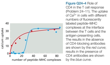 CHAPTER:Glossary CHAPTER:Index
-1 CD4 proteins on helper and regulatory T cells serve as co-receptors that bind to invariant parts of class II MHC proteins. CD4 is thought to increase the adhesion between T cells and antigen-presenting cells (APCs) that are initially connected only weakly by the T cell receptor bound to its specific peptide-MHC complex. To test this possibility, you label cell-surface MHC molecules with a fluorescently labeled peptide so that you can detect indi- vidual peptide-MHC complexes at the interface between the APCs and the T cells in a culture dish. To detect T cell responses-the sign of a productive contact-you load them with a Ca²+ indicator dye, as cytosolic Ca²+ increases when lymphocytes are active. You now count the peptide- MHC complexes at a large number of interfaces (immuno- logical synapses) and measure the resulting uptake of Ca²+ in the adherent T cells (Figure Q24-4, red circles). When you repeat the experiment in the presence of blocking anti- bodies against CD4, you get a different result (blue circles). Do these results support or refute the notion that CD4 aug- ments T cell receptor binding? Explain your answer.
CHAPTER:Glossary CHAPTER:IndexExplanation Verified
The thymus forms a specialized organ of ...
Molecular Biology Of The Cell 6th Edition by Bruce Alberts, Alexander Johnson, Julian Lewis, David Morgan, Martin Raff, Keith Roberts, Peter Walter
Why don’t you like this exercise?
Other Minimum 8 character and maximum 255 character
Character 255


