Deck 8: Atherosclerosis
Full screen (f)
Question
A high power view of an arterial wall is shown in the accompanying photomicrograph.In the portion of the field labelled "FC",which one of the following cells is concentrated in this area? 
A)Fibroblasts
B)Intimal smooth muscle cells
C)Lymphocytes
D)Macrophages
E)Neutrophils
A)Fibroblasts
B)Intimal smooth muscle cells
C)Lymphocytes
D)Macrophages
E)Neutrophils
Question
A high power view of an arterial wall is shown in the accompanying photomicrograph.The area labelled "T" is best described by which one of the following words or phrases? 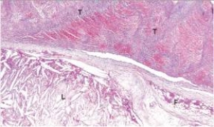
A)Acute inflammation
B)Cholesterol crystals
C)Fibrofatty plaque
D)Intimal proliferation
E)Thrombus
A)Acute inflammation
B)Cholesterol crystals
C)Fibrofatty plaque
D)Intimal proliferation
E)Thrombus
Question
In the accompanying photomicrograph of a small artery,the abnormality labelled "IP" refers to which one of the following? 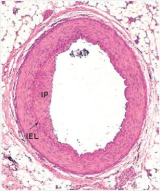
A)Iconographic proliferation
B)Idiopathic protein
C)Initial peninsula
D)Intimal proliferation
E)Ischemic portion
A)Iconographic proliferation
B)Idiopathic protein
C)Initial peninsula
D)Intimal proliferation
E)Ischemic portion
Question
A high power view of an arterial wall is shown in the accompanying photomicrograph.Which one of the following words or statements is the best classification of the atheromatous plaque shown? 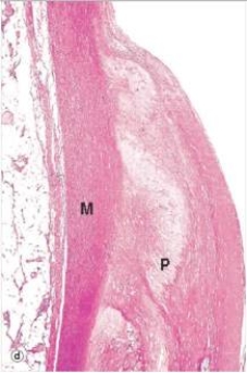
A)Fatty streak
B)Fibrofatty plaque
C)Fibrous plaque
D)Intimal proliferation
E)Necrotic plaque
A)Fatty streak
B)Fibrofatty plaque
C)Fibrous plaque
D)Intimal proliferation
E)Necrotic plaque
Question
The accompanying photomicrograph shows narrowing of an artery by an atheroma (A).Which one of the following would be the likely clinical symptom(s)if the artery in question were located in the lower extremities? 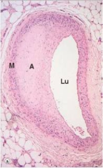
A)Muscle cramping
B)Claudication
C)Hemiplegia (paralysis of one leg)
D)Spasticity of the muscle
E)Tingling in the skin
A)Muscle cramping
B)Claudication
C)Hemiplegia (paralysis of one leg)
D)Spasticity of the muscle
E)Tingling in the skin
Question
A high power view of an arterial wall is shown in the accompanying photomicrograph.In the portion of the field labelled "C",which one of the following is responsible for the elongated,empty spaces seen? 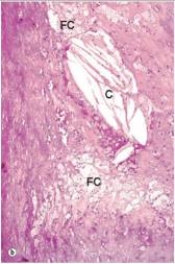
A)Coagulative necrosis
B)Cholesterol crystals
C)Liquifactive necrosis
D)Technical error
E)Uric acid crystals
A)Coagulative necrosis
B)Cholesterol crystals
C)Liquifactive necrosis
D)Technical error
E)Uric acid crystals
Question
The accompanying photomicrograph shows narrowing of an artery by an atheroma (A).Which one of the following would be the likely clinical symptom(s)if the artery in question were a coronary artery? 
A)Angina pectoris
B)Cramping of heart muscle
C)Myocardial infarction
D)Tingling in the skin over the heart
E)Ventricular fibrillation
A)Angina pectoris
B)Cramping of heart muscle
C)Myocardial infarction
D)Tingling in the skin over the heart
E)Ventricular fibrillation
Question
A high power view of an arterial wall is shown in the accompanying photomicrograph.Which one of the following words or statements is the best classification of the atheromatous plaque shown? 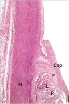
A)Fatty streak
B)Fibrofatty plaque
C)Fibrous plaque
D)Intimal proliferation
E)Necrotic plaque
A)Fatty streak
B)Fibrofatty plaque
C)Fibrous plaque
D)Intimal proliferation
E)Necrotic plaque

Unlock Deck
Sign up to unlock the cards in this deck!
Unlock Deck
Unlock Deck
1/8
Play
Full screen (f)
Deck 8: Atherosclerosis
1
A high power view of an arterial wall is shown in the accompanying photomicrograph.In the portion of the field labelled "FC",which one of the following cells is concentrated in this area?
A)Fibroblasts
B)Intimal smooth muscle cells
C)Lymphocytes
D)Macrophages
E)Neutrophils
A)Fibroblasts
B)Intimal smooth muscle cells
C)Lymphocytes
D)Macrophages
E)Neutrophils
Macrophages
2
A high power view of an arterial wall is shown in the accompanying photomicrograph.The area labelled "T" is best described by which one of the following words or phrases?
A)Acute inflammation
B)Cholesterol crystals
C)Fibrofatty plaque
D)Intimal proliferation
E)Thrombus
A)Acute inflammation
B)Cholesterol crystals
C)Fibrofatty plaque
D)Intimal proliferation
E)Thrombus
Thrombus
3
In the accompanying photomicrograph of a small artery,the abnormality labelled "IP" refers to which one of the following?
A)Iconographic proliferation
B)Idiopathic protein
C)Initial peninsula
D)Intimal proliferation
E)Ischemic portion
A)Iconographic proliferation
B)Idiopathic protein
C)Initial peninsula
D)Intimal proliferation
E)Ischemic portion
Intimal proliferation
4
A high power view of an arterial wall is shown in the accompanying photomicrograph.Which one of the following words or statements is the best classification of the atheromatous plaque shown?
A)Fatty streak
B)Fibrofatty plaque
C)Fibrous plaque
D)Intimal proliferation
E)Necrotic plaque
A)Fatty streak
B)Fibrofatty plaque
C)Fibrous plaque
D)Intimal proliferation
E)Necrotic plaque
Unlock Deck
Unlock for access to all 8 flashcards in this deck.
Unlock Deck
k this deck
5
The accompanying photomicrograph shows narrowing of an artery by an atheroma (A).Which one of the following would be the likely clinical symptom(s)if the artery in question were located in the lower extremities?
A)Muscle cramping
B)Claudication
C)Hemiplegia (paralysis of one leg)
D)Spasticity of the muscle
E)Tingling in the skin
A)Muscle cramping
B)Claudication
C)Hemiplegia (paralysis of one leg)
D)Spasticity of the muscle
E)Tingling in the skin
Unlock Deck
Unlock for access to all 8 flashcards in this deck.
Unlock Deck
k this deck
6
A high power view of an arterial wall is shown in the accompanying photomicrograph.In the portion of the field labelled "C",which one of the following is responsible for the elongated,empty spaces seen?
A)Coagulative necrosis
B)Cholesterol crystals
C)Liquifactive necrosis
D)Technical error
E)Uric acid crystals
A)Coagulative necrosis
B)Cholesterol crystals
C)Liquifactive necrosis
D)Technical error
E)Uric acid crystals
Unlock Deck
Unlock for access to all 8 flashcards in this deck.
Unlock Deck
k this deck
7
The accompanying photomicrograph shows narrowing of an artery by an atheroma (A).Which one of the following would be the likely clinical symptom(s)if the artery in question were a coronary artery?
A)Angina pectoris
B)Cramping of heart muscle
C)Myocardial infarction
D)Tingling in the skin over the heart
E)Ventricular fibrillation
A)Angina pectoris
B)Cramping of heart muscle
C)Myocardial infarction
D)Tingling in the skin over the heart
E)Ventricular fibrillation
Unlock Deck
Unlock for access to all 8 flashcards in this deck.
Unlock Deck
k this deck
8
A high power view of an arterial wall is shown in the accompanying photomicrograph.Which one of the following words or statements is the best classification of the atheromatous plaque shown?
A)Fatty streak
B)Fibrofatty plaque
C)Fibrous plaque
D)Intimal proliferation
E)Necrotic plaque
A)Fatty streak
B)Fibrofatty plaque
C)Fibrous plaque
D)Intimal proliferation
E)Necrotic plaque
Unlock Deck
Unlock for access to all 8 flashcards in this deck.
Unlock Deck
k this deck
Unlock Deck
Unlock for access to all 8 flashcards in this deck.


