Exam 11: Cranium
Exam 1: Preliminary Steps in Radiography32 Questions
Exam 2: General Anatomy and Radiographic Positioning Terminology119 Questions
Exam 3: Thoracic Viscera: Chest and Upper Airway79 Questions
Exam 4: Abdomen25 Questions
Exam 5: Upper Extremity136 Questions
Exam 6: Shoulder Girdle94 Questions
Exam 7: Lower Extremity154 Questions
Exam 8: Pelvis and Hip53 Questions
Exam 9: Vertebral Column179 Questions
Exam 10: Bony Thorax59 Questions
Exam 11: Cranium185 Questions
Exam 12: Trauma Radiography11 Questions
Exam 13: Contrast Arthrography3 Questions
Exam 14: Myelography and Other Central Nervous System Imaging13 Questions
Exam 15: Digestive System: Salivary Glands, alimentary Canal, and Biliary System125 Questions
Exam 16: Urinary System and Venipuncture81 Questions
Exam 17: Reproductive System9 Questions
Exam 18: Mammography27 Questions
Exam 19: Bone Densitometry16 Questions
Exam 20: Mobile Radiography12 Questions
Exam 21: Surgical Radiography11 Questions
Exam 22: Pediatric Imaging16 Questions
Exam 23: Geriatric Radiography14 Questions
Exam 24: Sectional Anatomy for Radiographers29 Questions
Exam 25: Computed Tomography14 Questions
Exam 26: Magnetic Resonance Imaging14 Questions
Exam 27: Vascular,cardiac,and Interventional Radiography37 Questions
Exam 28: Diagnostic Medical Sonography15 Questions
Exam 29: Nuclear Medicine and Molecular Imaging18 Questions
Exam 30: Radiation Oncology17 Questions
Select questions type
The figure below demonstrates the _____ method. 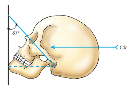
Free
(Multiple Choice)
4.8/5  (34)
(34)
Correct Answer: Verified
C
For a lateral projection of the facial bones,the image receptor is centered to the:
Free
(Multiple Choice)
4.8/5 (29)
Correct Answer:Verified
D
What is the average central-ray angulation for the PA axial (Haas)projection of the skull?
Free
(Multiple Choice)
4.7/5 (28)
Correct Answer:Verified
B
Which of the following reference lines is placed perpendicular to the image receptor for a parietoacanthial (Waters method)projection?
(Multiple Choice)
4.8/5 (32)
Letter A in the image below,used to evaluate the cranium,labels the: 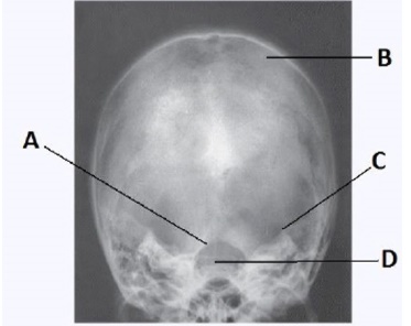
(Multiple Choice)
4.8/5 (43)
What projection (method)is demonstrated in the image below,used to evaluate the cranium? 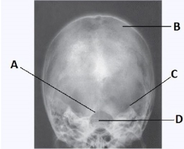
(Multiple Choice)
4.9/5 (32)
The OML forms an angle of how many degrees from the plane of the image receptor for an open-mouth parietoacanthial (Waters method)projection?
(Multiple Choice)
4.8/5 (40)
What is the central-ray angulation for the AP axial projection of the TMJ?
(Multiple Choice)
4.9/5 (36)
Which method of examining the skull is identified in the figure below? 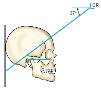
(Multiple Choice)
4.8/5 (36)
What is the central-ray angulation for the AP axial projection of the zygomatic arches if OML is used as the positioning baseline?
(Multiple Choice)
4.9/5 (33)
Which two parts of the patient's face touch the table for a PA axial projection (Caldwell method)? (Select all that apply.)
(Multiple Choice)
4.8/5 (30)
The part of the sphenoid bone identified in the figure below is the: 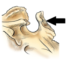
(Multiple Choice)
4.8/5 (38)
Which bone in the skull contains the auditory organs and the organs of hearing?
(Multiple Choice)
4.9/5 (33)
Which part of the patient's face is touching the upright bucky,or table,for a parietoacanthial projection (Waters method)?
(Multiple Choice)
4.8/5 (34)
The topographic line identified in the figure below is the: 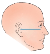
(Multiple Choice)
5.0/5 (41)
For the tangential projection of the zygomatic arch,the top of the head is tilted how many degrees?
(Multiple Choice)
4.8/5 (28)
Filters
- Essay(0)
- Multiple Choice(0)
- Short Answer(0)
- True False(0)
- Matching(0)