Exam 20: The Cardiovascular System: Blood
Exam 1: An Introduction to Anatomy110 Questions
Exam 2: The Cell110 Questions
Exam 3: The Tissue Level of Organization110 Questions
Exam 4: The Integumentary System110 Questions
Exam 5: The Skeletal System: Osseous Tissue and Skeletal Structure109 Questions
Exam 6: The Skeletal System: Axial Division109 Questions
Exam 7: The Skeletal System: Appendicular Division110 Questions
Exam 8: The Skeletal System: Articulations110 Questions
Exam 9: The Muscular System: Skeletal Muscle Tissue and Muscle Organization110 Questions
Exam 10: The Muscular System: the Axial Musculature110 Questions
Exam 11: The Muscular System: the Appendicular Musculature201 Questions
Exam 12: Surface Anatomy110 Questions
Exam 13: The Nervous System: Neural Tissue110 Questions
Exam 14: The Nervous System: the Spinal Cord and Spinal Nerves110 Questions
Exam 15: The Nervous System: the Brain and Cranial Nerves113 Questions
Exam 16: The Nervous System: Pathways and Higher-Order Functions117 Questions
Exam 17: The Nervous System: Autonomic Division117 Questions
Exam 18: The Nervous System: General and Special Senses116 Questions
Exam 19: The Endocrine System117 Questions
Exam 20: The Cardiovascular System: Blood112 Questions
Exam 21: The Cardiovascular System: the Heart116 Questions
Exam 22: The Cardiovascular System: Vessels and Circulation110 Questions
Exam 23: The Lymphatic System119 Questions
Exam 24: The Respiratory System116 Questions
Exam 25: The Digestive System122 Questions
Select questions type
Define and explain the significance of vascular anastomoses.
Free
(Essay)
4.8/5  (44)
(44)
Correct Answer: Verified
A vascular anastomosis is a ʺcoming togetherʺ of either arteries or veins. Vascular anastomoses are alternative pathways for blood flow. These pathways are called collateral channels. Arterial anastomoses often occur around joints providing alternative pathways for blood to flow when movement of a joint impinges upon flow through other vessels. The anastomosis ensures adequate perfusion of the tissue.
Venous anastomoses provide multiple pathways for drainage. Blockage of a single vein rarely blocks blood flow or leads to tissue death.
The pulse can be palpated near the temple anterior to the auricle of the ear.
Free
(Short Answer)
4.9/5 (35)
Correct Answer:Verified
temporal
Which vessel is missing in the following statement? ʺTracing venous blood from the inferior left side of the posterior abdominal wall to the heart, we find that blood enters the posterior intercostal veins, the hemiazygos vein, the superior vena cava, and the right atrium.ʺ
Free
(Multiple Choice)
4.7/5 (39)
Correct Answer:Verified
B
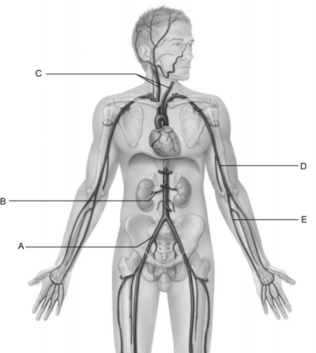 Figure 20.2
Use the diagram above to answer the following questions.
-Identify the letter that indicates the renal artery.
Figure 20.2
Use the diagram above to answer the following questions.
-Identify the letter that indicates the renal artery.
(Multiple Choice)
4.8/5 (33)
In metabolically active tissues, blood is present in metarterioles, and precapillary sphincters are constricted.
(True/False)
4.9/5 (44)
Systemic blood pressure is regulated by adjusting the diameter of arterioles.
(True/False)
4.7/5 (39)
What artery enters the skull through the foramen spinosum and supplies the inner surface of the parietal bone, dura mater, and parts of the temporal bone?
(Multiple Choice)
4.8/5 (36)
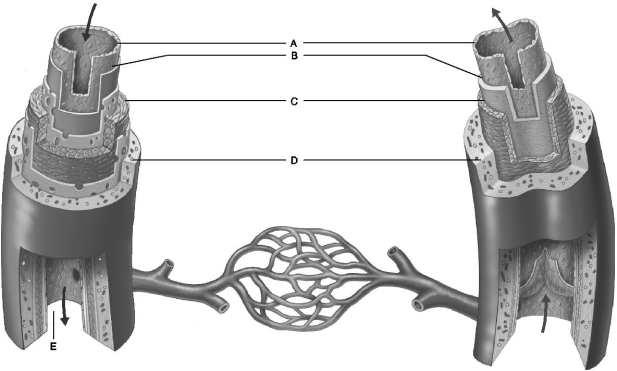 Figure 20.1
Use the diagram above to answer the following questions.
-Identify the letter that indicates a connective tissue layer consisting of longitudinal collagen fibers.
Figure 20.1
Use the diagram above to answer the following questions.
-Identify the letter that indicates a connective tissue layer consisting of longitudinal collagen fibers.
(Multiple Choice)
4.9/5 (29)
Which vessel is missing from the following statement? ʺTracing blood that drains from the large intestine, we find that blood drains from the distal colon is collected in the inferior mesenteric vein, merges with the splenic vein then directed to the hepatic portal vein, the liver sinusoids, and the inferior vena cava.ʺ
(Multiple Choice)
4.9/5 (41)
The extensor muscles of the forearm are supplied by which artery?
(Multiple Choice)
5.0/5 (33)
A preferred site to insert intravenous catheters is into the
(Multiple Choice)
4.8/5 (35)
Vessels of the small intestines, renal glomerulus, and synovial membranes that allow passage of fluid and solutes through ʺwindowsʺ in the endothelium.
(Multiple Choice)
4.8/5 (35)
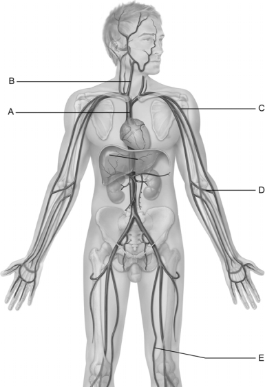 Figure 20.3
Use the diagram above to answer the following questions.
-Identify the letter that indicates the vessel that arises from the union of the left and right brachiocephalic veins.
Figure 20.3
Use the diagram above to answer the following questions.
-Identify the letter that indicates the vessel that arises from the union of the left and right brachiocephalic veins.
(Multiple Choice)
4.8/5 (52)
Venous blood from the right posterior intercostal veins (except the first intercostal space) flows to the unpaired vein and on to the superior vena cava.
(Short Answer)
4.8/5 (35)
The largest blood vessels near the heart have their own blood supply network called the
.
(Short Answer)
4.9/5 (43)
The vein descends through the transverse foramina of the first six cervical vertebrae.
(Short Answer)
4.8/5 (44)
Two large (wide) arteries that have relatively superficial locations and are often wounded are the
(Multiple Choice)
4.8/5 (32)
The artery descends along the arcuate line of the ilium and passes below the inguinal ligament.
(Short Answer)
4.9/5 (40)
Figure 20.2
Use the diagram above to answer the following questions.
-Identify the letter that indicates the ulnar artery.
(Multiple Choice)
4.8/5 (31)
Filters
- Essay(0)
- Multiple Choice(0)
- Short Answer(0)
- True False(0)
- Matching(0)