Exam 7: Pelvis and Upper Femora
Exam 1: Preliminary Steps in Radiography33 Questions
Exam 2: Compensating Filters14 Questions
Exam 3: General Anatomy and Radiographic Positioning Terminology101 Questions
Exam 4: Upper Limb125 Questions
Exam 5: Shoulder Girdle92 Questions
Exam 6: Lower Limb143 Questions
Exam 7: Pelvis and Upper Femora53 Questions
Exam 8: Vertebral Column175 Questions
Exam 9: Bony Thorax58 Questions
Exam 10: Thoracic Viscera74 Questions
Exam 11: Long Bone Measurement10 Questions
Exam 12: Contrast Arthrography5 Questions
Exam 13: Trauma Radiography10 Questions
Exam 14: Mouth and Salivary Glands10 Questions
Exam 15: Anterior Part of Neck9 Questions
Exam 16: Abdomen30 Questions
Exam 17: Digestive System: Alimentary Canal130 Questions
Exam 18: Urinary System and Venipuncture96 Questions
Exam 19: Reproductive System11 Questions
Exam 20: Skull, Facial Bones, and Paranasal Sinuses196 Questions
Exam 21: Mammography35 Questions
Exam 22: Central Nervous System15 Questions
Exam 23: Vascular, Cardiac, and Interventional Radiography40 Questions
Exam 24: Pediatric Imaging20 Questions
Exam 25: Geriatric Radiography18 Questions
Exam 26: Mobile Radiography20 Questions
Exam 27: Surgical Radiography10 Questions
Exam 28: Sectional Anatomy for Radiographers30 Questions
Exam 29: Computed Tomography16 Questions
Exam 30: Magnetic Resonance Imaging21 Questions
Exam 31: Diagnostic Ultrasound14 Questions
Exam 32: Nuclear Medicine18 Questions
Exam 33: Bone Densitometry18 Questions
Exam 34: Radiation Oncology20 Questions
Select questions type
How much is the central ray angled for the AP oblique projection (Judet method) of the acetabulum?
(Multiple Choice)
4.9/5  (34)
(34)
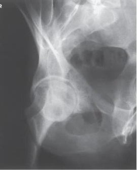
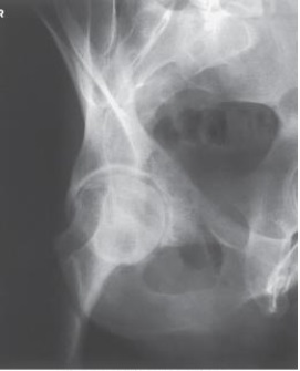
Examine this AP oblique (Judet) image of the right hip obtained with the patient positioned for the internal oblique. What patient position is depicted?
(Multiple Choice)
4.7/5 (38)
Examine this AP oblique (Judet) image of the right hip obtained with the patient positioned for the internal oblique. What is the anatomy of interest?
(Multiple Choice)
4.8/5 (40)
Which of the following describes the direction of the central ray for an axiolateral projection of the hip (Danelius-Miller)?
1)Perpendicular to the IR
2)Perpendicular to the long axis of the femoral neck
3)Perpendicular to the long axis of the femur
(Multiple Choice)
4.9/5 (41)
Flattening of the femoral head due to a vascular interruption is known as:
(Multiple Choice)
4.8/5 (43)
The body is placed at what angle for the AP oblique projection (Judet method) of the acetabulum?
(Multiple Choice)
4.7/5 (32)
To accurately position the patient for hip radiographs, one must localize two bony points on the pelvis. These two reference points are the:
1)superior margin of the symphysis.
2)greater trochanter of the femur.
3)anterior superior iliac spine.
(Multiple Choice)
4.8/5 (40)
The AP axial projection (Bridgeman method) requires that the central ray be directed:
(Multiple Choice)
4.8/5 (37)
In the anatomic position, the body of the femur is angled _____ degrees.
(Multiple Choice)
4.9/5 (28)
The respiration phase for the axiolateral projection of the hip (Danelius-Miller) is:
(Multiple Choice)
4.8/5 (37)
Which of the following projections can be performed with one exposure if a compensating filter is used?
(Multiple Choice)
4.8/5 (38)
Filters
- Essay(0)
- Multiple Choice(0)
- Short Answer(0)
- True False(0)
- Matching(0)