Deck 9: T-Cell-Mediated Immunity
Full screen (f)
Question
Question
Question
Question
Question
Question
Question
Question
Question
Question
A T cell line growing in culture is subjected to a chemical mutagen, and individual mutant lines are derived from this population. The individual mutant cell lines are each screened for their ability to proliferate in response to stimulation with antibodies to the T-cell receptor plus CD28 (anti-CD3 + anti-CD28). In addition, the cells are treated with varying doses of added IL-2, and three days later, T cell proliferation is measured by 3H-thymidine incorporation (cpm). The data for one mutant line and the wild-type control are shown in Figure Q16). 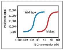
Figure Q16) The gene that is defective in this mutant T cell line most likely encodes:
A) The CD3 epsilon subunit of the T-cell receptor complex
B) The co-stimulatory molecule CD28
C) CD25, also known as the IL-2 receptor chain
D) The IL-2 receptor chain
E) The cytokine IL-2
Figure Q16) The gene that is defective in this mutant T cell line most likely encodes:
A) The CD3 epsilon subunit of the T-cell receptor complex
B) The co-stimulatory molecule CD28
C) CD25, also known as the IL-2 receptor chain
D) The IL-2 receptor chain
E) The cytokine IL-2
Question
Question
Question
Question
Question
Question
Question
Question
Question
Question
Naive T cells are isolated and left untreated or treated with 'compound X' for 1 hour. Following this, the T cells are incubated with a range of concentrations of a soluble form of ICAM-1 that has been conjugated to a fluorescent dye (soluble-ICAM-1-FITC). Fifteen minutes later, the cells are washed, and the relative amount of fluorescence bound to the cells is measured. The results of this assay are shown in Figure Q6). 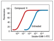 Figure Q6) The most likely identity of compound X is:
A) The adhesion molecule, LFA-1
B) The adhesion molecule, L-selectin
C) The sulfated carbohydrate structure, sulfated sialyl-LewisX
D) The chemokine ligand for CCR7
E) The immunoglobulin superfamily member, CD2
Figure Q6) The most likely identity of compound X is:A) The adhesion molecule, LFA-1
B) The adhesion molecule, L-selectin
C) The sulfated carbohydrate structure, sulfated sialyl-LewisX
D) The chemokine ligand for CCR7
E) The immunoglobulin superfamily member, CD2
Question
Question
Question
Question
Question
Question
Most effector T cells migrate out of secondary lymphoid organs and into tissues to exert their function. In which of the cases shown in Figure will the TH1 effector cell undergo long-lived interactions with its target cell, an infected macrophage? Assume all of the target cells shown below are infected with the pathogen recognized by the specific TH1 cells.
A)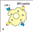
B)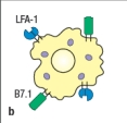
C)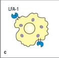
D)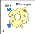
E)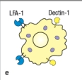
An immunological synapse forms between effector T cells and their targets to regulate signaling and to direct the release of effector molecules
A)
B)
C)
D)
E)
An immunological synapse forms between effector T cells and their targets to regulate signaling and to direct the release of effector molecules
Question
Question
Question
Question
Question
Question
Question
A mouse line (called 'Wm') is discovered that has an unexpected immunodeficiency. Genetic studies indicate that the immunodeficiency is due to a single gene defect. The Wm mice have normal numbers of all immune cell lineages, including T cell, B cells, macrophages, dendritic cells, NK cells, and granulocytes. When infected with viruses or intracellular bacteria and protozoa, the Wm mice mount normal protective T cell responses and clear the infections. However, given a helminthic parasite infection, the Wm mice cannot clear the infection and succumb to the disease. A set of wild-type (WT) and Wm mice were infected with the helminthic parasite, Nippostrongylus brasiliensis, and cytokine production by the CD4 T cells was analyzed. For this experiment, CD4 T cells were isolated from mice at day 8 post-infection, stimulated in vitro for 24 hr stimulation with anti-CD3 antibody to elicit cytokine secretion, and then cytokine levels in the culture supernatants were examined by ELISA. The results are shown in Figure.
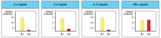
a) What process is likely defective in the Wm mice?
b) Which molecules are the best candidates for the defective gene in Wm mice? Note that the transcription factor GATA-3 is not a likely candidate gene, as GATA-3-deficient mice have an early block in T cell development in the thymus, and completely lack all T cells.and Wm mice and were stimulated in vitro with anti-CD3 plus anti-CD28 antibodies, in the presence of additional cytokines and blocking antibodies as indicated. After three days of stimulation, the T cells are isolated, washed, and restimulated with anti-CD3 antibody in medium lacking any cytokines or antibodies. After 24 hr, the cytokines present in the supernatants of these restimulated CD4 T cells are examined by ELISA. These data are show in Figure.
c) Do these results change your answers to parts (a) or (b) above?
To continue to identify the molecule or pathway defective in Wm T cells, CD4 T cells are isolated from Wm mice and stimulated in vitro with anti-CD3 + anti-CD28 antibodies in the presence of IL-4 and anti-IFN- antibody, as above. One day later, the cells are transduced with a vector expressing the transcription factor GATA3. From this vector, GATA3 will be expressed constitutively and at high levels, in all of the T cells. As a control, an 'empty' vector (not containing GATA-3) is also transduced into Wm CD4 T cells. Three days later, the cells are washed and restimulated for cytokine secretion analysis by ELISA, as above. Additionally, flow cytometry analysis of naive wild-type and Wm CD4 T cells stained with an antibody to the IL-4 receptor was performed, and the results are shown.and Wm CD4 T cells was then performed to examine the expression of the four Jak-family tyrosine kinases required for downstream signaling induced by the cytokine receptors expressed on T cells. The results from these experiments are shown in Figure.
d) With the new information provided by the data shown above, name the most likely candidate molecule for the defect in Wm T cells, and explain your reasoning.
Question
Question
Question
Question
West Nile virus (WNV) is a human pathogen that is transmitted by mosquitoes, which inject the virus into the host while they are feeding. To study the immune response to WNV, mice can be infected by injecting 100 PFU of virus into the footpad. This generates a robust immune response 7 days post-infection in the popliteal lymph node (LN), located behind the knee, as indicated in Figure.
Popliteal LN cells are isolated from the mouse shown above at day 7 post-infection, and the T cells are stimulated in vitro with the WNV peptide P33 plus dendritic cells isolated from a non-infected mouse of the same strain. Five hours later, the CD8 T cells in the culture are analyzed for their ability to produce the cytokine IFN- , and the numbers of IFN- -producing CD8 T cells are quantified. As a control, T cells are also stimulated with an irrelevant non-viral peptide (ova) plus dendritic cells. The results are shown in Figure.
a) Why is the T cell response different between the two lymph node populations?
To identify the important antigen-presenting cell required to activate WNV-specific CD8 T cells in the popliteal LN, wild-type mice and Batf3-/- mice are each immunized with 100 PFU of WNV in the left footpad, as above. Previous studies have indicated that Batf3-/- mice lack one particular subset of conventional dendritic cells, known as CD8 + dendritic cells (DC), but otherwise appear to have normal numbers and subsets of all other immune cell populations (e.g., T cells, B cells, macrophages, etc.). The results of this experiment are shown in Figure
b) Name two possible functions of CD8 + dendritic cells that could account for the results seen in the Batf3-/- mice immunized with WNV.
To distinguish between the possible functions of CD8 + dendritic cells, another set of wild-type and Batf3-/- mice are immunized with 100 PFU of WNV in the left footpad. At day 7 post-infection, the left popliteal LN are isolated from the mice, and the CD4 T cells in these LN populations are tested for responses to a WNV peptide bound to MHC class II (peptide Y). As a negative control, an MHC class II-binding peptide from ova is used. These results are shown in Figure .
c) Do these results rule in/out either of your proposed functions of CD8 + dendritic cells indicated in answer to part (b)? Why or why not?

Unlock Deck
Sign up to unlock the cards in this deck!
Unlock Deck
Unlock Deck
1/37
Play
Full screen (f)
Deck 9: T-Cell-Mediated Immunity
1
An immunodeficient mouse strain is identified, that has a single gene defect causing its disease. Mice with this defect have greatly impaired responses to protein antigens following subcutaneous immunization and also exhibit severely delays in the kinetics of their antibody responses. Analysis of their lymph nodes revealed profound alterations in the normal architecture, with a lack of organization of distinct T-cell and B-cell zones. A likely candidate for the defect in these mice is:
A) The chemokine receptor CCR7, which recruits B cells, T cells, and dendritic cells to lymph nodes.
B) The TNF family member LT- , which is made by Lti cells.
C) The chemokine receptor CXCR5 that recruits B cells to the lymph node follicles.
D) Prox1, the transcription factor required for the development of lymphatic vessels.
E) TNF- , which is required for the development of FDCs.
The chemokine receptor CCR7, which recruits B cells, T cells, and dendritic cells to lymph nodes.
2
To generate a vaccine to pertussis toxin, a heat-killed preparation of Bordetella pertussis (the bacteria that produce pertussis toxin) is mixed with a purified preparation of the inactivated toxin protein. The mixture is then injected subcutaneously into mice. In an effort to enhance the antibody response to the toxin, a group of test mice is depleted of all their dendritic cells immediately prior to immunization. However, instead of enhancing the antibody response, the dendritic cell depletion nearly eliminated the anti-toxin antibody response because:
A) All of the toxin-specific B cells underwent apoptosis in the absence of dendritic cells.
B) The heat-killed Bordetella pertussis could not be ingested by macrophages in the mice.
C) Dendritic cells are needed to activate naive CD4 T cells, which can then help the B cells produce anti-toxin antibody.
D) The macrophages in the mice did not express the appropriate pattern recognition receptors to recognize Bordetella pertussis.
E) The toxin-specific B cells did not up-regulate their MHC class II molecules after encountering the Bordetella pertussis.
Dendritic cells are needed to activate naive CD4 T cells, which can then help the B cells produce anti-toxin antibody.
3
Naive T cells scan the dendritic cells in the cortical region of the lymph node as they migrate. The initial encounter of T cells with dendritic cells is mediated by interactions between the T-cell receptor and the peptide:MHC complexes on the dendritic cell.
False
4
While CD28 co-stimulation is important for the initial activation of naive T cells, other co-stimulatory molecules function at later stages of the T cell response. Several of these other co-stimulatory molecules are members of the TNF-receptor family, and function by activating the transcription factor, NF B. Therefore, stimulation of these co-stimulatory TNF-receptors on activated T cells is likely to:
A) Induce programmed cell death in the T cell
B) Enhance T cell survival
C) Increase T cell proliferation
D) Promote T cell migration into tissues
E) Inhibit T cell activation and differentiation
A) Induce programmed cell death in the T cell
B) Enhance T cell survival
C) Increase T cell proliferation
D) Promote T cell migration into tissues
E) Inhibit T cell activation and differentiation
Unlock Deck
Unlock for access to all 37 flashcards in this deck.
Unlock Deck
k this deck
5
When T cells are activated by recognizing peptide:MHC complexes on dendritic cells in the lymph node, they up-regulate the receptor CD69. For T cells expressing a given T-cell receptor, the initial strength of the T-cell receptor signal can be modulated by varying the number of peptide:MHC complexes on the dendritic cells, or by varying the affinity with which the T cell-receptor binds to the peptide:MHC complexes. As a result, T cells stimulated with stronger T-cell receptor signals will maintain high expression of CD69 for one or two days longer that if those same T cells were stimulated with weaker T-cell receptor signals. Therefore, T cells stimulated with weaker T-cell receptor signals are likely to:
A) Die by apoptosis
B) Undergo more rounds of proliferation that T cells stimulated with stronger T-cell receptor signals
C) Migrate to the B-cell zones of the lymph node
D) Have reduced effector functions, such as cytokine production
E) Egress from the lymph node 1-2 days earlier than T cells stimulated with stronger T-cell receptor signals
Unlock Deck
Unlock for access to all 37 flashcards in this deck.
Unlock Deck
k this deck
6
Inbred strains of mice often generate highly polarized CD4 T cell responses to specific infections that are dominated by one subset of effector cells. In the case of Balb/c mice infected with the intracellular protozoan Leishmania major, a robust CD4 T cell response is elicited, generating large numbers of L. major-specific T cells; however, this response does not eliminate the pathogen, and instead the mice succumb to the infection. One likely explanation for this finding is:
A) L. major produces immune evasion molecules that prevent pathogen clearance.
B) The antibody response to L. major is too weak to eradicate the pathogen.
C) The CD4 T cell response produces effector cytokines that do not activate macrophages.
D) The CD4 T cells have differentiated into TH1 instead of TH2 effector cells.
E) The TFH cells specific for L. major induce a non-protective class of immunoglobulins.
Unlock Deck
Unlock for access to all 37 flashcards in this deck.
Unlock Deck
k this deck
7
The TNF family of cytokines and their receptors are critical for the development of secondary lymphoid organs, such as the lymph nodes and Peyer's patches. As a consequence, knockout mice lacking expression of LT- fail to develop most of these structures. Reconstitution of irradiated LT- -deficient mice with bone marrow stem cells from wild-type mice (e.g., LT- -sufficient) would:
A) Restore all missing lymphoid structures in the recipient mice
B) Restore the missing lymphoid structures but not the missing follicular dendritic cells in the recipient mice
C) Restore the missing follicular dendritic cells but not the missing lymphoid structures in the recipient mice
D) Have no effect on any lymphoid structures in the recipient mice
E) Only restore the proper organization of B cell follicles in the recipient mice
Unlock Deck
Unlock for access to all 37 flashcards in this deck.
Unlock Deck
k this deck
8
Secondary lymphoid organs, such as the lymph nodes, spleen, and mucosa-associated lymphoid tissues, each have distinct features that are important for their role in initiating immune responses focused on different anatomical compartments (i.e., the peripheral tissues, the blood, or the gastrointestinal tract, respectively). Yet these organs share some overall structural features, such as distinct T-cell and B-cell zones. One major difference between these organs is:
A) The presence versus the absence of macrophages and dendritic cells
B) Their function in bringing rare naive lymphocytes into contact with their specific antigen
C) The ability of lymphocytes to enter the organ from the blood
D) The mechanism by which antigens or pathogens enter the organ
E) The ability of naïve T cells to be activated and proliferate in the organ
Unlock Deck
Unlock for access to all 37 flashcards in this deck.
Unlock Deck
k this deck
9
When macrophages in the lung are infected with an intracellular bacterium, such as Mycobacterium tuberculosis, they are activated by the interactions of their pattern recognition receptors with the microbe. These activated macrophages will then:
A) Migrate to the marginal sinus of the draining lymph node to activate naive CD4 T cells
B) Increase their phagocytic properties to scavenge dead and dying neighboring cells
C) Migrate to the liver where they will remove dying cells from the blood
D) Migrate to the red pulp of the spleen where they will remove immune complexes from the circulation
E) Up-regulate MHC class II and B7 molecules to amplify CD4 T cell responses in the lung
Unlock Deck
Unlock for access to all 37 flashcards in this deck.
Unlock Deck
k this deck
10
A T cell line growing in culture is subjected to a chemical mutagen, and individual mutant lines are derived from this population. The individual mutant cell lines are each screened for their ability to proliferate in response to stimulation with antibodies to the T-cell receptor plus CD28 (anti-CD3 + anti-CD28). In addition, the cells are treated with varying doses of added IL-2, and three days later, T cell proliferation is measured by 3H-thymidine incorporation (cpm). The data for one mutant line and the wild-type control are shown in Figure Q16).
Figure Q16) The gene that is defective in this mutant T cell line most likely encodes:
A) The CD3 epsilon subunit of the T-cell receptor complex
B) The co-stimulatory molecule CD28
C) CD25, also known as the IL-2 receptor chain
D) The IL-2 receptor chain
E) The cytokine IL-2
Figure Q16) The gene that is defective in this mutant T cell line most likely encodes:
A) The CD3 epsilon subunit of the T-cell receptor complex
B) The co-stimulatory molecule CD28
C) CD25, also known as the IL-2 receptor chain
D) The IL-2 receptor chain
E) The cytokine IL-2
Unlock Deck
Unlock for access to all 37 flashcards in this deck.
Unlock Deck
k this deck
11
The entry of naive T cells from the blood into lymph nodes and mucosal lymphoid tissues occurs by a process that involves similar steps and similar adhesion molecules to the process by which leukocytes are recruited into sites of inflammation. Yet naive T cells do not enter tissues at sites of inflammation, but rather, home to lymphoid tissues. Which class of adhesion molecules direct the specific homing of naive T cells to lymphoid tissues?
Unlock Deck
Unlock for access to all 37 flashcards in this deck.
Unlock Deck
k this deck
12
Purified naive T cells isolated from a T-cell receptor transgenic mouse represent a homogeneous population of cells with specificity for a single known peptide:MHC complex. This specific peptide:MHC complex can be purified and formed into multivalent complexes, such as peptide:MHC tetramers. When the naive T cells are stimulated with their 'antigen' in the form of these peptide:MHC tetramers, the T cells show activation responses, including the up-regulation of genes that are induced within several hours after T-cell receptor stimulation. However, these activated T cells fail to undergo robust proliferation, and the majority of cells die after 3-4 days in culture. T cell proliferation and survival could be greatly augmented by addition of:
A) Anti-inflammatory cytokines
B) Toll-like receptor ligands
C) Antibodies to the T-cell receptor
D) B7 ligands for CD28
E) The integrin LFA-1
A) Anti-inflammatory cytokines
B) Toll-like receptor ligands
C) Antibodies to the T-cell receptor
D) B7 ligands for CD28
E) The integrin LFA-1
Unlock Deck
Unlock for access to all 37 flashcards in this deck.
Unlock Deck
k this deck
13
Chimeric mice are generated where approximately 50% of the cells in the animal are genetically MHC class I-deficient. The other 50% are deficient for the herpes virus receptor, HVEM, but do express MHC class I molecules. When these mice are infected with herpesvirus by intraperitoneal injection, a robust virus-specific CD8 T cell response is detected at day 7 post-infection in the spleens of the infected mice. Which cells are presenting herpesvirus antigens to prime the CD8 T cell response, and how did they acquire the viral antigens? Microbe-induced TLR signaling in tissue-resident dendritic cells induces their migration to lymphoid organs and enhances antigen processing
Unlock Deck
Unlock for access to all 37 flashcards in this deck.
Unlock Deck
k this deck
14
A mouse is infected with staphylococcal bacteria through a laceration in the skin of its paw. Dendritic cells are isolated from the tissue at the site of infection, and are incubated together with naïve staphylococcal-specific CD4 T cells. Seventy-two hours later, the proliferation of the CD4 T cells is measured as a readout for T cell activation. Surprisingly, the T cell response is quite poor compared to the response observed when the same T cells are mixed with a comparable number of dendritic cells isolated from the draining lymph node of the infected mouse. A comparison of the dendritic cells isolated from the two different sites would reveal:
A) Much higher levels of MHC and B7 molecules on the lymph node dendritic cells than those from the infected tissue
B) Much higher expression of all TLRs in the lymph node dendritic cells than those from the infected tissue
C) An increased number of MHC class II molecules bearing bacterial peptides on the surface of dendritic cells from the infected tissue than on those from the lymph node
D) Increased phagocytic activity of the lymph node dendritic cells than those from the infected tissue
E) Increased expression of Dectin-1, DEC205, and DC-SIGN on the lymph node dendritic cells than on those from the infected tissue
Unlock Deck
Unlock for access to all 37 flashcards in this deck.
Unlock Deck
k this deck
15
Many virus infections induce robust production of type I interferons, IFN- and IFN- . One example is the herpes simplex virus-1 (HSV-2), one of the most common sexually transmitted diseases in the US. Experiments that investigated the immune response to HSV-2 using a mouse model demonstrated essential roles for the DNA sensing Toll-like receptor, TLR9, and its downstream signaling adapter, MyD88, in the production of type I interferons following HSV-2 immunization. The type I interferon response to HSV-2 would be restored in Tlr9-/- mice by transferring into these mice wild-type:
A) Macrophages
B) Conventional dendritic cells
C) Mast cells
D) Plasmacytoid dendritic cells
E) Neutrophils
A) Macrophages
B) Conventional dendritic cells
C) Mast cells
D) Plasmacytoid dendritic cells
E) Neutrophils
Unlock Deck
Unlock for access to all 37 flashcards in this deck.
Unlock Deck
k this deck
16
A mouse is immunized with a single 9 amino acid peptide derived from the influenza virus. This peptide binds to MHC class I and produces an epitope (peptide:MHC complex) recognized by a small number of naive CD8 T cells in the mouse. The peptide is mixed with CpG oligonucleotides that are ligands for TLR-9. Surprisingly, this immunization regimen generates a very poor cytotoxic CD8 effector response to targets coated with this peptide compared to immunization with a preparation of intact heat-killed influenza virus mixed with CpG oligonucleotides. The enhanced cytotoxic T cell response to the peptide observed following immunization with intact viral particles compared to the peptide alone is due to:
A) The presence of CD4 T cell epitopes in the intact virus
B) The increased production of type I interferon elicited by the intact virus
C) The presence of additional CD8 epitopes in the intact virus
D) Presentation of peptides by macrophages instead of dendritic cells
E) Up-regulation of MHC class I molecules by the intact virus
Unlock Deck
Unlock for access to all 37 flashcards in this deck.
Unlock Deck
k this deck
17
Unlike innate immune responses, adaptive immune responses are initiated in secondary lymphoid organs. However, the innate immune response to an infection in a tissue has a pivotal role in inducing T-cell responses in the nearest lymph node by activating tissue dendritic cells and inducing their migration to the lymph node.
Unlock Deck
Unlock for access to all 37 flashcards in this deck.
Unlock Deck
k this deck
18
Strep throat is commonly caused by group A Streptococcus bacteria. A common symptom of strep throat is the presence of swollen lymph nodes in the neck. This symptom usually peaks about 2-4 days after the onset of the infection, and is due to:
A) Damage to the pharyngeal epithelium by the bacteria
B) Release of bacterial PAMPs leading to inflammatory cytokine production
C) Trapping and activation of antigen-specific lymphocytes in the lymph nodes of the neck
D) Recruitment of neutrophils to the lymph nodes of the neck
E) Recruitment of circulating macrophages to the lymph nodes of the neck
Unlock Deck
Unlock for access to all 37 flashcards in this deck.
Unlock Deck
k this deck
19
To avoid the potential for activating self-reactive T cells that might cause autoimmunity, naive T cell activation has stringent requirements for co-stimulatory signals in addition to the engagement of T-cell receptors with peptide:MHC complexes. Yet this requirement is abandoned once T cells have differentiated into effector cells. Name two effector T cell functions that would fail if effector T cells also required T-cell receptor signals plus co-stimulatory signals through CD28 to elicit their effector responses.
Unlock Deck
Unlock for access to all 37 flashcards in this deck.
Unlock Deck
k this deck
20
Naive T cells are isolated and left untreated or treated with 'compound X' for 1 hour. Following this, the T cells are incubated with a range of concentrations of a soluble form of ICAM-1 that has been conjugated to a fluorescent dye (soluble-ICAM-1-FITC). Fifteen minutes later, the cells are washed, and the relative amount of fluorescence bound to the cells is measured. The results of this assay are shown in Figure Q6). Figure Q6) The most likely identity of compound X is:
A) The adhesion molecule, LFA-1
B) The adhesion molecule, L-selectin
C) The sulfated carbohydrate structure, sulfated sialyl-LewisX
D) The chemokine ligand for CCR7
E) The immunoglobulin superfamily member, CD2
Figure Q6) The most likely identity of compound X is:A) The adhesion molecule, LFA-1
B) The adhesion molecule, L-selectin
C) The sulfated carbohydrate structure, sulfated sialyl-LewisX
D) The chemokine ligand for CCR7
E) The immunoglobulin superfamily member, CD2
Unlock Deck
Unlock for access to all 37 flashcards in this deck.
Unlock Deck
k this deck
21
Several effector T cell functions are mediated by cell surface molecules on the effector cell interacting with binding partners on the target cells. In the case of the CD40-CD40 ligand interaction, effector CD4 T cells express CD40 ligand, which binds to CD40 target cells. Individuals with a genetic deficiency in CD40 ligand expression show greatly reduced antibody responses, particularly to protein antigens which require CD4 TFH cell interactions with B cells. Another expected defect in these individuals would be:
A) Reduced recruitment of eosinophils by TH2 cells
B) Reduced recruitment of neutrophils by TH17 cells
C) Reduced production of anti-microbial peptides induced by TH17 cells
D) Reduced suppression of dendritic cells by Treg cells
E) Reduced activation of macrophages by TH1 cells
A) Reduced recruitment of eosinophils by TH2 cells
B) Reduced recruitment of neutrophils by TH17 cells
C) Reduced production of anti-microbial peptides induced by TH17 cells
D) Reduced suppression of dendritic cells by Treg cells
E) Reduced activation of macrophages by TH1 cells
Unlock Deck
Unlock for access to all 37 flashcards in this deck.
Unlock Deck
k this deck
22
In a lymph node, nTreg cells are able to inhibit the responses of other T cells in their vicinity. This inhibition is specific, as the nTreg cell and the naive T cell must share the same antigen specificity.
Unlock Deck
Unlock for access to all 37 flashcards in this deck.
Unlock Deck
k this deck
23
Cytotoxic granules released from cytotoxic T cells contain proteins that enter target cells and induce apoptosis. Do any of the mechanisms of apoptosis induction by cytotoxic effector T cells rely on the protein APAF-1? If so, which pathway?
Unlock Deck
Unlock for access to all 37 flashcards in this deck.
Unlock Deck
k this deck
24
Eosinophils are a subset of granulocytes that normally reside in the circulation. When activated, these cells secrete toxic compounds that are a key component in the eradication of helminthic parasite infections. Eosinophils are recruited to sites of parasite infections by:
A) Interferon- produced by TH1 cells
B) CCL11 produced by TH2 cells
C) Interferon- produced by CD8 cytotoxic T cells
D) CCL20 produced by TH17 cells
E) GM-CSF produced by TH1, TH2, and TH17 cells
A) Interferon- produced by TH1 cells
B) CCL11 produced by TH2 cells
C) Interferon- produced by CD8 cytotoxic T cells
D) CCL20 produced by TH17 cells
E) GM-CSF produced by TH1, TH2, and TH17 cells
Unlock Deck
Unlock for access to all 37 flashcards in this deck.
Unlock Deck
k this deck
25
Each subset of effector T cells produces a distinct array of secreted factors known as cytokines. One common feature of nearly all effector T cell responses is the rapid production of increased numbers of macrophages, granulocytes, and dendritic cells from the bone marrow. This response occurs due to:
A) Effector T cell trafficking to the bone marrow to activate hematopoietic progenitor cells
B) Depletion of peripheral myeloid cells after leaving the blood, leading to increased bone marrow production
C) Inflammation-induced blood vessel dilation causing transient reductions in circulating myeloid cells
D) Pathogen-induced migration of tissue-resident dendritic cells to lymph nodes inducing new dendritic cell production
E) Effector T cell production of GM-CSF that traffics to the bone marrow
Unlock Deck
Unlock for access to all 37 flashcards in this deck.
Unlock Deck
k this deck
26
Most effector T cells migrate out of secondary lymphoid organs and into tissues to exert their function. In which of the cases shown in Figure will the TH1 effector cell undergo long-lived interactions with its target cell, an infected macrophage? Assume all of the target cells shown below are infected with the pathogen recognized by the specific TH1 cells.
A)
B)
C)
D)
E)
An immunological synapse forms between effector T cells and their targets to regulate signaling and to direct the release of effector molecules
A)
B)
C)
D)
E)
An immunological synapse forms between effector T cells and their targets to regulate signaling and to direct the release of effector molecules
Unlock Deck
Unlock for access to all 37 flashcards in this deck.
Unlock Deck
k this deck
27
Cytotoxic T cells that lack expression of perforin are more defective in killing target cells than those that lack granzymes.
Unlock Deck
Unlock for access to all 37 flashcards in this deck.
Unlock Deck
k this deck
28
The compound LE135 is an inhibitor of the retinoic acid receptor, and blocks signaling through this receptor. In a mouse model of inflammatory bowel disease (IBD), inflammation in the gastrointestinal (GI) epithelium is significantly exacerbated if animals are treated with LE135. Analysis of the CD4 T cell subsets found in the GI epithelium of LE135-treated mice compared to controls with IBD would likely show:
A) Reduced numbers of CD4 T cells of all subsets in the LE135-treated mice
B) Increased numbers of TGF- and IL-10-producing CD4 cells in the LE135-treated mice
C) Reduced numbers of IL-17 and IL-22-producing CD4 cells in the LE135-treated mice
D) Reduced numbers of iTregs in the LE135-treated mice
E) Increased numbers of naive CD4 T cells in the LE135-treated mice
Unlock Deck
Unlock for access to all 37 flashcards in this deck.
Unlock Deck
k this deck
29
The Bcl-2 protein was first identified based on its overexpression in a subset of B cell lymphomas, where it was shown to promote the resistance of the tumor cells to apoptosis. Subcellular localization experiments would show that Bcl-2 is present:
A) In the nucleus, where it blocks DNA fragmentation
B) In the cytotoxic granules where it inactivates granzymes
C) In the mitochondria where it blocks cytochrome c release
D) In the cytosol where it blocks caspase activation
E) In the plasma membrane where it blocks death receptor signaling
A) In the nucleus, where it blocks DNA fragmentation
B) In the cytotoxic granules where it inactivates granzymes
C) In the mitochondria where it blocks cytochrome c release
D) In the cytosol where it blocks caspase activation
E) In the plasma membrane where it blocks death receptor signaling
Unlock Deck
Unlock for access to all 37 flashcards in this deck.
Unlock Deck
k this deck
30
Cytotoxic T cells are rapid killers of infected target cells. Within minutes of the interaction of a cytotoxic T cell with a target cell, the program of apoptosis in the target cell is initiated. This rapid activity is a consequence of:
A) The expression and packaging of perforin and granzymes in cytotoxic granules prior to target cell encounter
B) The extremely rapid production of granzymes and perforin by cytotoxic effector cells upon encountering a target cell
C) The extremely potent activity of perforins in poking holes in target cell membranes allowing entry of granzymes
D) The efficient activity of granzymes in cleaving effector caspases leading to direct activation of apoptosis
E) The large number of cytotoxic granules made and secreted by a single effector cytotoxic T cell
Unlock Deck
Unlock for access to all 37 flashcards in this deck.
Unlock Deck
k this deck
31
Wiskott-Aldrich syndrome is an immunodeficiency disease that causes a defect in antibody responses. The most severe defects are in antibody responses to protein antigens, which are dependent on CD4 effector TFH cells providing cytokines to the B cell. The protein defective in individuals with the disease, known as WASp, functions in cytoskeletal reorganization and polarization through its role in promoting actin polymerization. This immune defect could be fixed by a gene therapy approach that restored WASp expression in:
A) Antigen-presenting cells such as dendritic cells
B) B cells
C) B cells and T cells
D) T cells
E) T cells and antigen-presenting cells
A) Antigen-presenting cells such as dendritic cells
B) B cells
C) B cells and T cells
D) T cells
E) T cells and antigen-presenting cells
Unlock Deck
Unlock for access to all 37 flashcards in this deck.
Unlock Deck
k this deck
32
Cytotoxic effector T cells also produce inflammatory cytokines such as IFN- and TNF- when their T-cell receptor recognizes peptide:MHC on a target cell. One effect of this cytokine secretion is to enhance the ability of CD8 effector T cells to recognize and kill other infected cells in the nearby vicinity. This enhanced activity is due to:
A) The increased production of perforin and granzymes by CD8 cells
B) The up-regulation of MHC class I protein expression by IFN-
C) The ability of TNF- to induce vascular leakage
D) The effect of cytokines on promoting target cell apoptosis
E) The effect of IFN- to enhance viral replication leading to increased viral antigen presentation
Unlock Deck
Unlock for access to all 37 flashcards in this deck.
Unlock Deck
k this deck
33
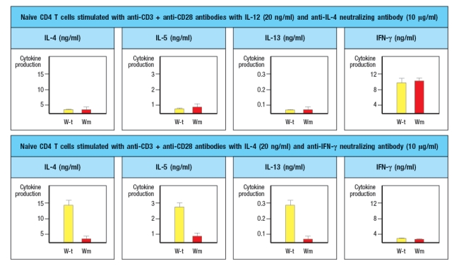
A mouse line (called 'Wm') is discovered that has an unexpected immunodeficiency. Genetic studies indicate that the immunodeficiency is due to a single gene defect. The Wm mice have normal numbers of all immune cell lineages, including T cell, B cells, macrophages, dendritic cells, NK cells, and granulocytes. When infected with viruses or intracellular bacteria and protozoa, the Wm mice mount normal protective T cell responses and clear the infections. However, given a helminthic parasite infection, the Wm mice cannot clear the infection and succumb to the disease. A set of wild-type (WT) and Wm mice were infected with the helminthic parasite, Nippostrongylus brasiliensis, and cytokine production by the CD4 T cells was analyzed. For this experiment, CD4 T cells were isolated from mice at day 8 post-infection, stimulated in vitro for 24 hr stimulation with anti-CD3 antibody to elicit cytokine secretion, and then cytokine levels in the culture supernatants were examined by ELISA. The results are shown in Figure.
a) What process is likely defective in the Wm mice?
b) Which molecules are the best candidates for the defective gene in Wm mice? Note that the transcription factor GATA-3 is not a likely candidate gene, as GATA-3-deficient mice have an early block in T cell development in the thymus, and completely lack all T cells.and Wm mice and were stimulated in vitro with anti-CD3 plus anti-CD28 antibodies, in the presence of additional cytokines and blocking antibodies as indicated. After three days of stimulation, the T cells are isolated, washed, and restimulated with anti-CD3 antibody in medium lacking any cytokines or antibodies. After 24 hr, the cytokines present in the supernatants of these restimulated CD4 T cells are examined by ELISA. These data are show in Figure.
c) Do these results change your answers to parts (a) or (b) above?
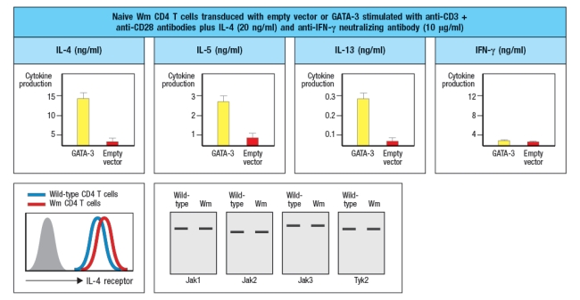
To continue to identify the molecule or pathway defective in Wm T cells, CD4 T cells are isolated from Wm mice and stimulated in vitro with anti-CD3 + anti-CD28 antibodies in the presence of IL-4 and anti-IFN- antibody, as above. One day later, the cells are transduced with a vector expressing the transcription factor GATA3. From this vector, GATA3 will be expressed constitutively and at high levels, in all of the T cells. As a control, an 'empty' vector (not containing GATA-3) is also transduced into Wm CD4 T cells. Three days later, the cells are washed and restimulated for cytokine secretion analysis by ELISA, as above. Additionally, flow cytometry analysis of naive wild-type and Wm CD4 T cells stained with an antibody to the IL-4 receptor was performed, and the results are shown.and Wm CD4 T cells was then performed to examine the expression of the four Jak-family tyrosine kinases required for downstream signaling induced by the cytokine receptors expressed on T cells. The results from these experiments are shown in Figure.
d) With the new information provided by the data shown above, name the most likely candidate molecule for the defect in Wm T cells, and explain your reasoning.
a) What process is likely defective in the Wm mice?
b) Which molecules are the best candidates for the defective gene in Wm mice? Note that the transcription factor GATA-3 is not a likely candidate gene, as GATA-3-deficient mice have an early block in T cell development in the thymus, and completely lack all T cells.and Wm mice and were stimulated in vitro with anti-CD3 plus anti-CD28 antibodies, in the presence of additional cytokines and blocking antibodies as indicated. After three days of stimulation, the T cells are isolated, washed, and restimulated with anti-CD3 antibody in medium lacking any cytokines or antibodies. After 24 hr, the cytokines present in the supernatants of these restimulated CD4 T cells are examined by ELISA. These data are show in Figure.
c) Do these results change your answers to parts (a) or (b) above?
To continue to identify the molecule or pathway defective in Wm T cells, CD4 T cells are isolated from Wm mice and stimulated in vitro with anti-CD3 + anti-CD28 antibodies in the presence of IL-4 and anti-IFN- antibody, as above. One day later, the cells are transduced with a vector expressing the transcription factor GATA3. From this vector, GATA3 will be expressed constitutively and at high levels, in all of the T cells. As a control, an 'empty' vector (not containing GATA-3) is also transduced into Wm CD4 T cells. Three days later, the cells are washed and restimulated for cytokine secretion analysis by ELISA, as above. Additionally, flow cytometry analysis of naive wild-type and Wm CD4 T cells stained with an antibody to the IL-4 receptor was performed, and the results are shown.and Wm CD4 T cells was then performed to examine the expression of the four Jak-family tyrosine kinases required for downstream signaling induced by the cytokine receptors expressed on T cells. The results from these experiments are shown in Figure.
d) With the new information provided by the data shown above, name the most likely candidate molecule for the defect in Wm T cells, and explain your reasoning.
Unlock Deck
Unlock for access to all 37 flashcards in this deck.
Unlock Deck
k this deck
34
At early timepoints following an infection, examination of lymph node CD4 T cells responding to the pathogen would show a heterogeneous population of cells representing several different effector lineages. Likewise, the cytokines produced by these cells would include IFN , IL-4, and possibly others. However, approximately one week later at the peak of the T cell response, the pathogen-specific CD4 T cell population would be largely homogeneous in their production of a single effector subset cytokine profile. This change comes about due to:
A) Death of the CD4 T cells making the non-protective cytokines
B) Increased proliferation of CD4 T cells making protective cytokines
C) Enhanced differentiation of newly activated CD4 T cells into one effector subset
D) Inhibition of antigen-presenting cells by CD4 T cell-derived cytokines
E) Impaired proliferation of effector CD4 T cells by IFN
Unlock Deck
Unlock for access to all 37 flashcards in this deck.
Unlock Deck
k this deck
35
Effector caspases are activated downstream of both extrinsic and intrinsic pathways of apoptosis. Consequently, cells lacking one or more of these enzymes show defects in apoptosis. An alternative means of eliminating the activity of an effector caspase would be to:
A) Generate a form of the pro-caspase with a mutation in the initiator caspase cleavage site
B) Express a mutant form the caspase that lacked the pro-domain
C) Express a constitutively active form of the initiator caspase
D) Generate a knockout mutation that prevented expression of the Fas receptor
E) Generate a mutation in the proteins required for autophagy
Unlock Deck
Unlock for access to all 37 flashcards in this deck.
Unlock Deck
k this deck
36
Following their activation, naive CD4 T cells differentiate into one of several subsets of effector T cells. This developmental choice is determined by which specific cytokines the T cells are exposed to during their activation; in turn, the cytokines that are produced will reflect the nature of the infecting pathogen. In order for this system to be flexible enough to produce all five effector CD4 subsets (TH1, TH2, TH17, TFH, and iTreg):
A) Each naive CD4 T cell must already express one of the lineage-specific transcription factors (i.e., T-bet, GATA-3, etc.).
B) Each naive CD4 T cell must express all of the STAT proteins.
C) Each naive CD4 T cell must express only a single STAT protein (i.e., STAT1 or STAT6 or STAT3, etc.).
D) Each naive CD4 T cell must already express low levels of the subset specific cytokines (i.e., IFN , IL-4, IL-17, etc.).
E) Each naive CD4 T cell must express all of the lineage-specific transcription factors (i.e., T-bet, GATA-3, etc.).
Unlock Deck
Unlock for access to all 37 flashcards in this deck.
Unlock Deck
k this deck
37
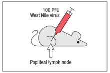
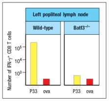
West Nile virus (WNV) is a human pathogen that is transmitted by mosquitoes, which inject the virus into the host while they are feeding. To study the immune response to WNV, mice can be infected by injecting 100 PFU of virus into the footpad. This generates a robust immune response 7 days post-infection in the popliteal lymph node (LN), located behind the knee, as indicated in Figure.
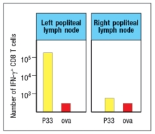
Popliteal LN cells are isolated from the mouse shown above at day 7 post-infection, and the T cells are stimulated in vitro with the WNV peptide P33 plus dendritic cells isolated from a non-infected mouse of the same strain. Five hours later, the CD8 T cells in the culture are analyzed for their ability to produce the cytokine IFN- , and the numbers of IFN- -producing CD8 T cells are quantified. As a control, T cells are also stimulated with an irrelevant non-viral peptide (ova) plus dendritic cells. The results are shown in Figure.
a) Why is the T cell response different between the two lymph node populations?
To identify the important antigen-presenting cell required to activate WNV-specific CD8 T cells in the popliteal LN, wild-type mice and Batf3-/- mice are each immunized with 100 PFU of WNV in the left footpad, as above. Previous studies have indicated that Batf3-/- mice lack one particular subset of conventional dendritic cells, known as CD8 + dendritic cells (DC), but otherwise appear to have normal numbers and subsets of all other immune cell populations (e.g., T cells, B cells, macrophages, etc.). The results of this experiment are shown in Figure
b) Name two possible functions of CD8 + dendritic cells that could account for the results seen in the Batf3-/- mice immunized with WNV.
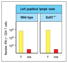
To distinguish between the possible functions of CD8 + dendritic cells, another set of wild-type and Batf3-/- mice are immunized with 100 PFU of WNV in the left footpad. At day 7 post-infection, the left popliteal LN are isolated from the mice, and the CD4 T cells in these LN populations are tested for responses to a WNV peptide bound to MHC class II (peptide Y). As a negative control, an MHC class II-binding peptide from ova is used. These results are shown in Figure .
c) Do these results rule in/out either of your proposed functions of CD8 + dendritic cells indicated in answer to part (b)? Why or why not?
Popliteal LN cells are isolated from the mouse shown above at day 7 post-infection, and the T cells are stimulated in vitro with the WNV peptide P33 plus dendritic cells isolated from a non-infected mouse of the same strain. Five hours later, the CD8 T cells in the culture are analyzed for their ability to produce the cytokine IFN- , and the numbers of IFN- -producing CD8 T cells are quantified. As a control, T cells are also stimulated with an irrelevant non-viral peptide (ova) plus dendritic cells. The results are shown in Figure.
a) Why is the T cell response different between the two lymph node populations?
To identify the important antigen-presenting cell required to activate WNV-specific CD8 T cells in the popliteal LN, wild-type mice and Batf3-/- mice are each immunized with 100 PFU of WNV in the left footpad, as above. Previous studies have indicated that Batf3-/- mice lack one particular subset of conventional dendritic cells, known as CD8 + dendritic cells (DC), but otherwise appear to have normal numbers and subsets of all other immune cell populations (e.g., T cells, B cells, macrophages, etc.). The results of this experiment are shown in Figure
b) Name two possible functions of CD8 + dendritic cells that could account for the results seen in the Batf3-/- mice immunized with WNV.
To distinguish between the possible functions of CD8 + dendritic cells, another set of wild-type and Batf3-/- mice are immunized with 100 PFU of WNV in the left footpad. At day 7 post-infection, the left popliteal LN are isolated from the mice, and the CD4 T cells in these LN populations are tested for responses to a WNV peptide bound to MHC class II (peptide Y). As a negative control, an MHC class II-binding peptide from ova is used. These results are shown in Figure .
c) Do these results rule in/out either of your proposed functions of CD8 + dendritic cells indicated in answer to part (b)? Why or why not?
Unlock Deck
Unlock for access to all 37 flashcards in this deck.
Unlock Deck
k this deck
Unlock Deck
Unlock for access to all 37 flashcards in this deck.


