Exam 9: T-Cell-Mediated Immunity
Exam 1: Basic Concepts in Immunology44 Questions
Exam 2: Innate Immunity: the First Lines of Defense32 Questions
Exam 3: The Induced Responses of Innate Immunity39 Questions
Exam 4: Antigen Recognition by B-Cell and T-Cell Receptors28 Questions
Exam 5: The Generation of Lymphocyte Antigen Receptors33 Questions
Exam 6: Antigen Presentation to T Lymphocytes30 Questions
Exam 7: Lymphocyte Receptor Signaling42 Questions
Exam 8: Development and Survival of Lymphocytes37 Questions
Exam 9: T-Cell-Mediated Immunity37 Questions
Exam 10: The Humoral Immune Response30 Questions
Exam 11: Integrated Dynamics of Innate and Adaptive Immunity28 Questions
Exam 12: The Mucosal Immune System27 Questions
Exam 13: Failures of Host Defense Mechanisms43 Questions
Exam 14: Allergy and Allergic Diseases26 Questions
Exam 15: Autoimmunity and Transplantation31 Questions
Exam 16: Manipulation of the Immune Response34 Questions
Select questions type
The Bcl-2 protein was first identified based on its overexpression in a subset of B cell lymphomas, where it was shown to promote the resistance of the tumor cells to apoptosis. Subcellular localization experiments would show that Bcl-2 is present:
Free
(Multiple Choice)
4.8/5  (26)
(26)
Correct Answer: Verified
C
While CD28 co-stimulation is important for the initial activation of naive T cells, other co-stimulatory molecules function at later stages of the T cell response. Several of these other co-stimulatory molecules are members of the TNF-receptor family, and function by activating the transcription factor, NF B. Therefore, stimulation of these co-stimulatory TNF-receptors on activated T cells is likely to:
Free
(Multiple Choice)
4.9/5 (28)
Correct Answer:Verified
B
Cytotoxic T cells that lack expression of perforin are more defective in killing target cells than those that lack granzymes.
Free
(True/False)
4.8/5 (24)
Correct Answer:Verified
True
Wiskott-Aldrich syndrome is an immunodeficiency disease that causes a defect in antibody responses. The most severe defects are in antibody responses to protein antigens, which are dependent on CD4 effector TFH cells providing cytokines to the B cell. The protein defective in individuals with the disease, known as WASp, functions in cytoskeletal reorganization and polarization through its role in promoting actin polymerization. This immune defect could be fixed by a gene therapy approach that restored WASp expression in:
(Multiple Choice)
4.8/5 (26)
Inbred strains of mice often generate highly polarized CD4 T cell responses to specific infections that are dominated by one subset of effector cells. In the case of Balb/c mice infected with the intracellular protozoan Leishmania major, a robust CD4 T cell response is elicited, generating large numbers of L. major-specific T cells; however, this response does not eliminate the pathogen, and instead the mice succumb to the infection. One likely explanation for this finding is:
(Multiple Choice)
4.8/5 (41)
Eosinophils are a subset of granulocytes that normally reside in the circulation. When activated, these cells secrete toxic compounds that are a key component in the eradication of helminthic parasite infections. Eosinophils are recruited to sites of parasite infections by:
(Multiple Choice)
4.9/5 (36)
The compound LE135 is an inhibitor of the retinoic acid receptor, and blocks signaling through this receptor. In a mouse model of inflammatory bowel disease (IBD), inflammation in the gastrointestinal (GI) epithelium is significantly exacerbated if animals are treated with LE135. Analysis of the CD4 T cell subsets found in the GI epithelium of LE135-treated mice compared to controls with IBD would likely show:
(Multiple Choice)
4.8/5 (37)
Many virus infections induce robust production of type I interferons, IFN- and IFN- . One example is the herpes simplex virus-1 (HSV-2), one of the most common sexually transmitted diseases in the US. Experiments that investigated the immune response to HSV-2 using a mouse model demonstrated essential roles for the DNA sensing Toll-like receptor, TLR9, and its downstream signaling adapter, MyD88, in the production of type I interferons following HSV-2 immunization. The type I interferon response to HSV-2 would be restored in Tlr9-/- mice by transferring into these mice wild-type:
(Multiple Choice)
4.8/5 (48)
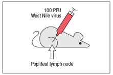
West Nile virus (WNV) is a human pathogen that is transmitted by mosquitoes, which inject the virus into the host while they are feeding. To study the immune response to WNV, mice can be infected by injecting 100 PFU of virus into the footpad. This generates a robust immune response 7 days post-infection in the popliteal lymph node (LN), located behind the knee, as indicated in Figure.
 Popliteal LN cells are isolated from the mouse shown above at day 7 post-infection, and the T cells are stimulated in vitro with the WNV peptide P33 plus dendritic cells isolated from a non-infected mouse of the same strain. Five hours later, the CD8 T cells in the culture are analyzed for their ability to produce the cytokine IFN- , and the numbers of IFN- -producing CD8 T cells are quantified. As a control, T cells are also stimulated with an irrelevant non-viral peptide (ova) plus dendritic cells. The results are shown in Figure.
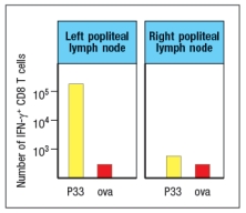 a) Why is the T cell response different between the two lymph node populations?
To identify the important antigen-presenting cell required to activate WNV-specific CD8 T cells in the popliteal LN, wild-type mice and Batf3-/- mice are each immunized with 100 PFU of WNV in the left footpad, as above. Previous studies have indicated that Batf3-/- mice lack one particular subset of conventional dendritic cells, known as CD8 + dendritic cells (DC), but otherwise appear to have normal numbers and subsets of all other immune cell populations (e.g., T cells, B cells, macrophages, etc.). The results of this experiment are shown in Figure
a) Why is the T cell response different between the two lymph node populations?
To identify the important antigen-presenting cell required to activate WNV-specific CD8 T cells in the popliteal LN, wild-type mice and Batf3-/- mice are each immunized with 100 PFU of WNV in the left footpad, as above. Previous studies have indicated that Batf3-/- mice lack one particular subset of conventional dendritic cells, known as CD8 + dendritic cells (DC), but otherwise appear to have normal numbers and subsets of all other immune cell populations (e.g., T cells, B cells, macrophages, etc.). The results of this experiment are shown in Figure
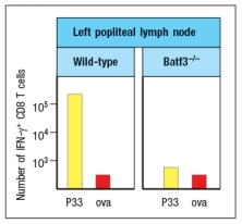 b) Name two possible functions of CD8 + dendritic cells that could account for the results seen in the Batf3-/- mice immunized with WNV.
To distinguish between the possible functions of CD8 + dendritic cells, another set of wild-type and Batf3-/- mice are immunized with 100 PFU of WNV in the left footpad. At day 7 post-infection, the left popliteal LN are isolated from the mice, and the CD4 T cells in these LN populations are tested for responses to a WNV peptide bound to MHC class II (peptide Y). As a negative control, an MHC class II-binding peptide from ova is used. These results are shown in Figure .
b) Name two possible functions of CD8 + dendritic cells that could account for the results seen in the Batf3-/- mice immunized with WNV.
To distinguish between the possible functions of CD8 + dendritic cells, another set of wild-type and Batf3-/- mice are immunized with 100 PFU of WNV in the left footpad. At day 7 post-infection, the left popliteal LN are isolated from the mice, and the CD4 T cells in these LN populations are tested for responses to a WNV peptide bound to MHC class II (peptide Y). As a negative control, an MHC class II-binding peptide from ova is used. These results are shown in Figure .
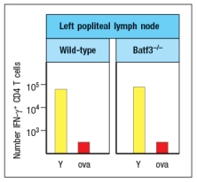 c) Do these results rule in/out either of your proposed functions of CD8 + dendritic cells indicated in answer to part (b)? Why or why not?
(Essay)
4.8/5 (28)
A mouse is infected with staphylococcal bacteria through a laceration in the skin of its paw. Dendritic cells are isolated from the tissue at the site of infection, and are incubated together with naïve staphylococcal-specific CD4 T cells. Seventy-two hours later, the proliferation of the CD4 T cells is measured as a readout for T cell activation. Surprisingly, the T cell response is quite poor compared to the response observed when the same T cells are mixed with a comparable number of dendritic cells isolated from the draining lymph node of the infected mouse. A comparison of the dendritic cells isolated from the two different sites would reveal:
(Multiple Choice)
4.9/5 (31)
In a lymph node, nTreg cells are able to inhibit the responses of other T cells in their vicinity. This inhibition is specific, as the nTreg cell and the naive T cell must share the same antigen specificity.
(True/False)
4.9/5 (18)
When T cells are activated by recognizing peptide:MHC complexes on dendritic cells in the lymph node, they up-regulate the receptor CD69. For T cells expressing a given T-cell receptor, the initial strength of the T-cell receptor signal can be modulated by varying the number of peptide:MHC complexes on the dendritic cells, or by varying the affinity with which the T cell-receptor binds to the peptide:MHC complexes. As a result, T cells stimulated with stronger T-cell receptor signals will maintain high expression of CD69 for one or two days longer that if those same T cells were stimulated with weaker T-cell receptor signals. Therefore, T cells stimulated with weaker T-cell receptor signals are likely to:
(Multiple Choice)
4.7/5 (37)
Each subset of effector T cells produces a distinct array of secreted factors known as cytokines. One common feature of nearly all effector T cell responses is the rapid production of increased numbers of macrophages, granulocytes, and dendritic cells from the bone marrow. This response occurs due to:
(Multiple Choice)
4.9/5 (29)
An immunodeficient mouse strain is identified, that has a single gene defect causing its disease. Mice with this defect have greatly impaired responses to protein antigens following subcutaneous immunization and also exhibit severely delays in the kinetics of their antibody responses. Analysis of their lymph nodes revealed profound alterations in the normal architecture, with a lack of organization of distinct T-cell and B-cell zones. A likely candidate for the defect in these mice is:
(Multiple Choice)
4.9/5 (33)
Secondary lymphoid organs, such as the lymph nodes, spleen, and mucosa-associated lymphoid tissues, each have distinct features that are important for their role in initiating immune responses focused on different anatomical compartments (i.e., the peripheral tissues, the blood, or the gastrointestinal tract, respectively). Yet these organs share some overall structural features, such as distinct T-cell and B-cell zones. One major difference between these organs is:
(Multiple Choice)
4.9/5 (37)
To avoid the potential for activating self-reactive T cells that might cause autoimmunity, naive T cell activation has stringent requirements for co-stimulatory signals in addition to the engagement of T-cell receptors with peptide:MHC complexes. Yet this requirement is abandoned once T cells have differentiated into effector cells. Name two effector T cell functions that would fail if effector T cells also required T-cell receptor signals plus co-stimulatory signals through CD28 to elicit their effector responses.
(Essay)
4.9/5 (30)
A mouse is immunized with a single 9 amino acid peptide derived from the influenza virus. This peptide binds to MHC class I and produces an epitope (peptide:MHC complex) recognized by a small number of naive CD8 T cells in the mouse. The peptide is mixed with CpG oligonucleotides that are ligands for TLR-9. Surprisingly, this immunization regimen generates a very poor cytotoxic CD8 effector response to targets coated with this peptide compared to immunization with a preparation of intact heat-killed influenza virus mixed with CpG oligonucleotides. The enhanced cytotoxic T cell response to the peptide observed following immunization with intact viral particles compared to the peptide alone is due to:
(Multiple Choice)
4.8/5 (38)
When macrophages in the lung are infected with an intracellular bacterium, such as Mycobacterium tuberculosis, they are activated by the interactions of their pattern recognition receptors with the microbe. These activated macrophages will then:
(Multiple Choice)
4.8/5 (27)
Several effector T cell functions are mediated by cell surface molecules on the effector cell interacting with binding partners on the target cells. In the case of the CD40-CD40 ligand interaction, effector CD4 T cells express CD40 ligand, which binds to CD40 target cells. Individuals with a genetic deficiency in CD40 ligand expression show greatly reduced antibody responses, particularly to protein antigens which require CD4 TFH cell interactions with B cells. Another expected defect in these individuals would be:
(Multiple Choice)
4.8/5 (28)
At early timepoints following an infection, examination of lymph node CD4 T cells responding to the pathogen would show a heterogeneous population of cells representing several different effector lineages. Likewise, the cytokines produced by these cells would include IFN , IL-4, and possibly others. However, approximately one week later at the peak of the T cell response, the pathogen-specific CD4 T cell population would be largely homogeneous in their production of a single effector subset cytokine profile. This change comes about due to:
(Multiple Choice)
4.8/5 (38)
Filters
- Essay(0)
- Multiple Choice(0)
- Short Answer(0)
- True False(0)
- Matching(0)