Deck 7: Lymphocyte Receptor Signaling
Full screen (f)
Question
Question
Question
Question
Question
Question
Question
Question
Question
Question
Question
Question
Question
Using an antibody that recognizes the phosphorylated, but not the non-phosphorylated form of the transcription factor, NFAT, T cells are permeabilized, stained with this antibody, and analyzed by flow cytometry. Which of the data in Figure represent the expected pattern of staining from wild-type T cells before and after TCR stimulation. 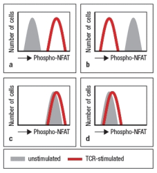
Question
Question
Question
Question
Question
Question
Question
Question
A mutant B cell line is examined by confocal microscopy after incubation with a microbial pathogen recognized by the BCR on these B cells. The B cells have been stained with antibodies to visualize the localization of polymerized actin and microtubules. As a control, wild-type B cells are examined. The results are shown in Figure, with the numbers indicating the proportion of cells examined that show each pattern of staining. 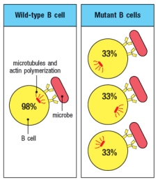
To identify the specific signaling defect in these mutant B cells, a reasonable biochemical assay would be to:
A) Determine if BCR stimulation of mutant B cells produces enhanced binding of the B cell to the microbe
B) Determine whether the mutant B cells have reduced levels of the enzyme Protein kinase C-
C) Determine whether the mutant B cells are overexpressing the enzyme Vav
D) Determine whether BCR stimulation of mutant B cells promotes exchange of GDP for GTP on cdc42
E) Determine whether BCR stimulation of mutant B cells produces increased levels of DAG
Question
Question
Antigen receptor signaling and lymphocyte activation.antibody coupled to biotin, followed by cross-linking with Streptavidin (S-Av). As the antibody and then S-Av are added, the cells are run on the flow cytometer to examine the fluorescence of the Ca2+-sensitive dye. After several minutes of analysis, the cells are stimulated with ionomycin (Iono), to induce Ca2+ influx; this is used as a positive control to ensure that the cells are loaded with the dye. In Figure Q41)A, the characteristic pattern of Ca2+ influx is shown in the red line (wild-type; WT), where TCR stimulation causes a sharp rise in cytoplasmic Ca2+, followed by a slow decline over hours. As shown below, cytoplasmic Ca2+ concentrations do not normally return to baseline for the timecourse of this experiment. A mutant mouse is identified with a defect in T cell activation in response to TCR stimulation. The calcium response of T cells from the mutant mouse is shown in the blue line. 
a) Given these data, name three T cell signaling proteins that could be defective in the mutant T cells. Then name three T cell signaling proteins that could not be responsible for this defect, even if mutated.
Additional experiments are performed to analyze protein tyrosine phosphorylation in response to TCR stimulation. For these experiments, T cells are stimulated with anti-CD3 antibody, and then lysates are prepared and run on a protein (SDS-PAGE) gel to separate the proteins by molecular weight. The proteins are transferred from the gel to a membrane for immunoblotting using an antibody that binds to all phosphorylated tyrosine residues in any protein; this antibody is called ‘anti-phospho-tyrosine antibody,’ and is abbreviated as anti-P-Y. The results are shown in Figure .
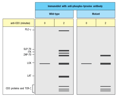
You confirm that the mutant T cells express normal levels of all the proteins detected in the WT cells, including PLC- , SLP-76, ITK, ZAP-70, LCK, LAT, and the CD3 and TCR proteins.
b) Based on these additional data, which of the candidate proteins in your answer to part (a) are ruled out? Briefly explain your answer.
c) What protein is most likely defective in the mutant cells and why?
d) For the protein you named in your answer to part (c), which amino acids or domain of the protein could be mutated to account for all the data.
a) Given these data, name three T cell signaling proteins that could be defective in the mutant T cells. Then name three T cell signaling proteins that could not be responsible for this defect, even if mutated.
Additional experiments are performed to analyze protein tyrosine phosphorylation in response to TCR stimulation. For these experiments, T cells are stimulated with anti-CD3 antibody, and then lysates are prepared and run on a protein (SDS-PAGE) gel to separate the proteins by molecular weight. The proteins are transferred from the gel to a membrane for immunoblotting using an antibody that binds to all phosphorylated tyrosine residues in any protein; this antibody is called ‘anti-phospho-tyrosine antibody,’ and is abbreviated as anti-P-Y. The results are shown in Figure .
You confirm that the mutant T cells express normal levels of all the proteins detected in the WT cells, including PLC- , SLP-76, ITK, ZAP-70, LCK, LAT, and the CD3 and TCR proteins.
b) Based on these additional data, which of the candidate proteins in your answer to part (a) are ruled out? Briefly explain your answer.
c) What protein is most likely defective in the mutant cells and why?
d) For the protein you named in your answer to part (c), which amino acids or domain of the protein could be mutated to account for all the data.
Question
T cells with defective TCR signaling are discovered, and found to have an inactivating mutation in a key TCR signaling protein. Using an antibody that recognizes the phosphorylated (activated) form of the Erk Map-kinase, stimulated T cells are permeabilized, stained with this antibody, and analyzed on the flow cytometer. These data are shown in Figure Q21). 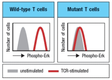 Figure Q21) Additional experiments examining Ca2+ influx into T cells following TCR stimulation show a normal response in the mutant T cells. One likely candidate gene that could be mutated in the defective cells is:
A) PLC-
B) ITK
C) RasGRP
D) Calcineurin
E) WASp
Figure Q21) Additional experiments examining Ca2+ influx into T cells following TCR stimulation show a normal response in the mutant T cells. One likely candidate gene that could be mutated in the defective cells is:A) PLC-
B) ITK
C) RasGRP
D) Calcineurin
E) WASp
Question
Question
Question
Question
Question
Question
Question
TCR stimulation was shown to affect ICAM-1 (integrin ligand) binding to LFA-1 (integrin) on T cells. To demonstrate this, varying concentrations of purified ICAM-1 were added to unstimulated or TCR-stimulated T cells, and the amount of ICAM-1 binding was measured. The data from such an experiment are displayed on Figure. Assign the red or blue lines correctly to 'unstimulated' or 'TCR-stimulated' T cells, and explain the reasoning for your answer. 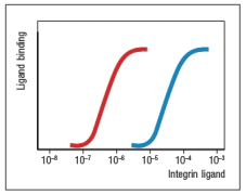
Question
Question
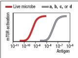
BCR signaling on B cells is initiated by antigen binding, leading to mTOR activation. This occurs, for instance, when the antigen is a live microbe that binds to the BCR on the B cells. Which one of the forms of antigen shown below the graph would correctly account for the data shown in Figure.
A)
B)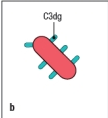
C)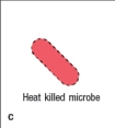
D)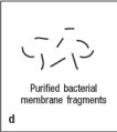
A)
B)
C)
D)
Question
Question
Question
Question
Question
Question
Question
Question
Synthesis question: Co-stimulatory and inhibitory receptors modulate antigen receptor signaling in T and B lymphocytes. A new receptor is discovered, expressed on the surface of T cells, and called 'X'. An antibody to X is generated, and used in T cell stimulation experiments. In these experiments, antibodies to the TCR complex (anti-CD3) and to CD28 (anti-CD28) are known to stimulate signaling through those receptors, as does the antibody to X. The data from an experiment measuring IL-2 secretion by the T cells stimulated with different combinations of antibodies are shown in Figure. 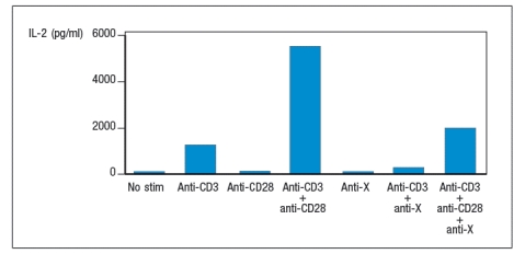
a) Does stimulation of receptor X alone induce IL-2 production by T cells? Does it enhance or inhibit TCR signaling? Indicate the evidence supporting your answers.
b) If you examined the amino acid sequence of the receptor X cytoplasmic tail, what motif would you expect to find?
Biochemical studies show that when receptor X is stimulated, a tyrosine residue in the cytoplasmic tail becomes phosphorylated.
c) From these data, what are the two most likely signaling proteins that might be recruited to receptor X when it is stimulated? Does the T cell stimulation data shown in the graph rule in or out either of your candidate proteins? Why or why not?
d) Describe a biochemical experiment (analysis of proteins) that would indicate which enzyme is recruited to receptor X when it is stimulated.
Question
Match between columns

Unlock Deck
Sign up to unlock the cards in this deck!
Unlock Deck
Unlock Deck
1/42
Play
Full screen (f)
Deck 7: Lymphocyte Receptor Signaling
1
Antigen receptors use multiple mechanisms to recruit signaling proteins to the plasma membrane, where they can propagate downstream signals. In some cases, recruitment of proteins to the membrane is induced following antigen receptor stimulation, whereas other proteins are constitutively associated with the membrane.
Name one mechanism that is induced by antigen receptor stimulation, and one that is constitutive, and give an example a protein recruited by each mechanism.
Name one mechanism that is induced by antigen receptor stimulation, and one that is constitutive, and give an example a protein recruited by each mechanism.
Recruitment induced by antigen receptor stimulation:
binding to a membrane associated scaffold protein that is phosphorylated in response to antigen receptor stimulation (examples: Grb2, Gads, SLP-76)
binding to the membrane phospholipid PIP3 that is generated by phosphorylation of PIP2 in response to antigen receptor stimulation (examples: Akt, Itk, PLC- )
Recruitment that is constitutive:
lipid modified proteins that associate constitutively with the plasma membrane (examples: small GTPases such as Ras, Rap1)
2
Antigen receptor signaling pathways are regulated by a balanced equilibrium between tyrosine kinases and tyrosine phosphatases. In general, activation of signaling proceeds when the kinase activities leading to auto-phosphorylation of Lck, to phosphorylation of ZAP-70, and to phosphorylation of downstream adapters and scaffolds exceeds the activity of phosphatases acting on these substrates. Therefore, it came as a surprise when T cells lacking the membrane tyrosine phosphatase, CD45, were first generated, and were found to be unable to be activated by TCR stimulation. Name one important function of CD45 in T cells that explains the requirement for this phosphatase in TCR signaling.
CD45 is the phosphatase that de-phosphorylates the C-terminal negative regulatory tyrosine of Lck. When this negative regulatory tyrosine is phosphorylated and binds to the Lck SH2 domain, Lck is held in an inactive conformation. TCR signaling initiated by Lck cannot occur without CD45 to dephosphorylate this site.
3
A new strain of immunodeficient mice has been discovered, and found to have T cells that are unresponsive to TCR stimulation. The T cells from these mice have normal levels of the TCR complex on their surface, but when this TCR is stimulated, the cells fail to secrete IL-2. As a first step in determining the signaling defect responsible for this immunodeficiency, the T cells are stimulated with a phorbol ester (PMA) and Ionomycin. It is found that this treatment elicits IL-2 production by the immunodeficient T cells. Based on this information, candidate genes that could be mutated in these T cells include all of the following EXCEPT:
A) ZAP-70
B) PLC-
C) SLP-76
D) ITK
E) Calcineurin
A) ZAP-70
B) PLC-
C) SLP-76
D) ITK
E) Calcineurin
Calcineurin
4
Many receptors of the immune system activate protein kinases as a mechanism of initiating signaling. For antigen receptors on lymphocytes, ligand binding induces receptor clustering, and the enzymes activated are protein tyrosine kinases. Based on this mechanism, predict the outcome of expressing a mutant form of the receptor-associated tyrosine kinase in cells that still express the wild-type version of this enzyme, and explain your reasoning. This mutant is unable to bind ATP and therefore is catalytically inactive; assume the mutant and wild-type forms of the kinase are expressed in equimolar amounts.
Unlock Deck
Unlock for access to all 42 flashcards in this deck.
Unlock Deck
k this deck
5
Antigen receptor signaling pathways are initiated by the action of a Src-family kinase. In T cells, the predominant Src-kinase is Lck. In resting T cells, Lck is maintained in an inactive state by allosteric interactions involving multiple domains of the enzyme. When T cells are treated with a small molecule inhibitor of the tyrosine kinase Csk, TCR signaling is initiated even in the absence of a ligand to stimulate the TCR. This occurs because:
A) Csk phosphorylates Lck in its kinase domain, leading to Lck activation.
B) Csk phosphorylates ZAP-70, maintaining ZAP-70 in an auto-inhibited state.
C) Csk phosphorylates the ITAM motifs in the TCR chain, leading to ZAP-70 recruitment.
D) Csk phosphorylates and activates the membrane tyrosine phosphatase CD45.
E) Csk phosphorylates the C-terminal negative regulatory tyrosine in Lck.
Unlock Deck
Unlock for access to all 42 flashcards in this deck.
Unlock Deck
k this deck
6
Immunoreceptor signaling proteins, such as the TCR chain and CD3 subunits, have conserved ITAM motifs in their cytoplasmic tails. When fully phosphorylated, the ITAM recruits a tyrosine kinase with a tandem SH2 domain structure at the amino-terminal end of the protein. Tandem SH2 domain-containing kinases do not bind to sequences in other proteins, even if they contain a phosphorylated tyrosine because:
A) The amino acid sequence adjacent to the phosphorylated tyrosines in the ITAM motif is unique, and not found in any other proteins.
B) The affinity of a single SH2 domain within these kinases for a tyrosine phosphorylated sequence is too low for efficient binding.
C) The amino-terminal SH2 domain of the kinase has very high affinity for both of the phosphorylated tyrosines in the ITAM motif, so will not bind to other proteins.
D) The amino-terminal SH2 domain of the kinase is in an autoinhibited conformation and can only bind to a phosphorylated ITAM.
E) The tandem SH2 domain-containing kinase phosphorylates the tyrosines in the ITAM itself, so can only bind to these sequences.
Unlock Deck
Unlock for access to all 42 flashcards in this deck.
Unlock Deck
k this deck
7
Following TCR stimulation, the small GTPase Ras is activated. Ras activation is induced by the Ras GTP-exchange factor (GEF), RasGRP. Both Ras and RasGRP are constitutively expressed in resting T cells. The reason Ras activation is only induced following TCR stimulation is:
A) RasGRP undergoes a Ca2+-dependent conformational change required for its activity.
B) RasGRP requires tyrosine phosphorylation for its activity.
C) RasGRP is ubiquitinated and degraded in the absence of TCR stimulation.
D) RasGRP recruitment to the plasma membrane requires TCR stimulation.
E) Ras is only recruited to the activated TCR following assembly of the LAT:Gads:SLP-76 complex.
Unlock Deck
Unlock for access to all 42 flashcards in this deck.
Unlock Deck
k this deck
8
The TCR signaling module leading to transcription factor activation is dependent on the enzyme phospholipase-C- (PLC- ). The mechanism by which PLC- activates multiple transcription factors is by:
A) Generating two small second messengers that act on multiple target proteins in the T cell
B) Directly cleaving inhibitory subunits of multiple transcription factors, thereby releasing the active transcription factors
C) Generating two small second messengers that diffuse to the nucleus and activate transcription factors present there
D) Generating two small second messengers that act as chaperones to promote nuclear localization of transcription factors
E) Directly cleaving the lipid binding domain from membrane-tethered transcription factors, allowing them to migrate to the nucleus
Unlock Deck
Unlock for access to all 42 flashcards in this deck.
Unlock Deck
k this deck
9
Small GTPases, such as Ras, Rho, and cdc42, are activated when they exchange their bound GDP for GTP. In the GTP-bound state, these proteins contribute to signaling by:
A) Hydrolyzing the bound GTP back to GDP
B) Interacting with GTPase-activating proteins (GAPs)
C) Interacting with target proteins and altering their activity
D) Diffusing from the membrane and entering the nucleus
E) Inducing calcium release from the endoplasmic reticulum
A) Hydrolyzing the bound GTP back to GDP
B) Interacting with GTPase-activating proteins (GAPs)
C) Interacting with target proteins and altering their activity
D) Diffusing from the membrane and entering the nucleus
E) Inducing calcium release from the endoplasmic reticulum
Unlock Deck
Unlock for access to all 42 flashcards in this deck.
Unlock Deck
k this deck
10
Several small GTPases play critical roles in antigen receptor signaling pathways. When activated by binding to GTP, these mediators induce changes in cytoskeletal organization, adhesion, and metabolism, but have no role in transcription factor activation.
Unlock Deck
Unlock for access to all 42 flashcards in this deck.
Unlock Deck
k this deck
11
The TCR and BCR are multi-subunit receptor complexes. Experiments examining the synthesis and transport of these receptors to the lymphocyte cell surface have shown that the signaling subunits of each receptor complex are required for transport of the ligand-binding receptor subunits to the cell surface. One possible reason for this stringent control on cell surface expression is:
A) To ensure that very few complete TCRs or BCRs are expressed on the lymphocyte surface
B) To ensure that each lymphocyte expresses only a single specificity of antigen receptor
C) To prevent surface expression of receptors that will bind ligand but fail to induce signals
D) To prevent lymphocytes from triggering antigen receptor signaling pathways from intracellular forms of the receptors
E) To ensure that equimolar amounts of all antigen receptor signaling subunits are produced
Unlock Deck
Unlock for access to all 42 flashcards in this deck.
Unlock Deck
k this deck
12
Second messengers, such as calcium ions (Ca2+), are chemical mediators commonly used in intracellular signaling pathways. Despite its common usage in many different cell types in the body, Ca2+ has specific effects in lymphocytes following antigen receptor stimulation. The specific responses of lymphocytes to increased concentrations of intracellular Ca2+ are determined by:
A) The expression of a specific subset of Ca2+-responsive enzymes in lymphocytes compared to other cell types
B) The increased expression of calmodulin in lymphocytes compared to other cell types
C) The presence of enzymes that bind calmodulin in lymphocytes but not other cell types
D) The high levels of Ca2+ in the endoplasmic reticulum of lymphocytes compared to other cell types
E) The ability of Ca2+ to amplify signaling pathways in lymphocytes but not other cell types
Unlock Deck
Unlock for access to all 42 flashcards in this deck.
Unlock Deck
k this deck
13
Using an antibody that recognizes the phosphorylated, but not the non-phosphorylated form of the transcription factor, NFAT, T cells are permeabilized, stained with this antibody, and analyzed by flow cytometry. Which of the data in Figure represent the expected pattern of staining from wild-type T cells before and after TCR stimulation.
Unlock Deck
Unlock for access to all 42 flashcards in this deck.
Unlock Deck
k this deck
14
Following TCR or BCR signaling, the most important events downstream of the activation of ZAP-70 or SYK, respectively, are the activation of transcription factors leading to new gene expression.
Unlock Deck
Unlock for access to all 42 flashcards in this deck.
Unlock Deck
k this deck
15
All of the modular protein domains used for signaling protein interactions bind to ligands that are transiently generated following receptor stimulation.
Unlock Deck
Unlock for access to all 42 flashcards in this deck.
Unlock Deck
k this deck
16
Human patients with genetic defects that result in a failure to produce the calcium channel protein ORAI1, or the ER calcium sensor protein STIM1, have severe immunodeficiency diseases. An immunosuppressive drug that would most closely mimic these primary immunodeficiencies is:
A) Rituximab, a drug that depletes B cells
B) Cyclosporin A, a calcineurin inhibitor
C) Rapamycin, an mTOR inhibitor
D) Tysabri, an inhibitor of integrin binding
E) Enbrel or Humira that inhibit TNF
A) Rituximab, a drug that depletes B cells
B) Cyclosporin A, a calcineurin inhibitor
C) Rapamycin, an mTOR inhibitor
D) Tysabri, an inhibitor of integrin binding
E) Enbrel or Humira that inhibit TNF
Unlock Deck
Unlock for access to all 42 flashcards in this deck.
Unlock Deck
k this deck
17
The TCR and BCR are each composed of two modules, an antigen-binding module and a signaling module; furthermore, in each case, the two functional modules are encoded by distinct polypeptides. In addition, the tyrosine kinases that initiate antigen receptor signaling are also separate proteins from those of each receptor. This is a different strategy for receptor signaling than the case of receptor tyrosine kinases, where the enzyme is an intrinsic component of the ligand-binding receptor protein. Name one advantage of this organization of the TCR and BCR that accounts for the expression of ZAP-70 and Syk, as well as ITAM-containing immunoreceptors, in many different subsets of immune cells.
Unlock Deck
Unlock for access to all 42 flashcards in this deck.
Unlock Deck
k this deck
18
Scaffold proteins are often phosphorylated at multiple sites, allowing the recruitment of several different signaling proteins. In antigen receptor signaling pathways, this mechanism is used to bring enzymes in close proximity to their substrates. Termination of this signaling mechanism would be most efficiently accomplished by:
A) Ubiquitination of the scaffold protein, leading to its degradation
B) Binding of the enzyme to a GTPase activating protein (GAP)
C) Depletion of the substrate due to enzyme catalysis
D) Dephosphorylation of the scaffold by a phosphatase
E) Ubiquitination of the enzyme by K63-linkage of polyubiquitin
Unlock Deck
Unlock for access to all 42 flashcards in this deck.
Unlock Deck
k this deck
19
Diacyl-glycerol (DAG) is one of the two products generated when PLC- cleaves the membrane phospholipid, PIP2. This small lipid mediator remains associated with the plasma membrane and functions to inhibit tyrosine phosphatases that remove activating phosphate groups from ZAP-70 and the Tec-family kinase, ITK.
Unlock Deck
Unlock for access to all 42 flashcards in this deck.
Unlock Deck
k this deck
20
The LAT:Gads:SLP-76 complex that assembles following TCR stimulation provides the scaffold for initiating multiple downstream signaling modules, leading to actin polymerization, integrin activation, and gene expression.
Unlock Deck
Unlock for access to all 42 flashcards in this deck.
Unlock Deck
k this deck
21
A mutant B cell line is examined by confocal microscopy after incubation with a microbial pathogen recognized by the BCR on these B cells. The B cells have been stained with antibodies to visualize the localization of polymerized actin and microtubules. As a control, wild-type B cells are examined. The results are shown in Figure, with the numbers indicating the proportion of cells examined that show each pattern of staining.
To identify the specific signaling defect in these mutant B cells, a reasonable biochemical assay would be to:
A) Determine if BCR stimulation of mutant B cells produces enhanced binding of the B cell to the microbe
B) Determine whether the mutant B cells have reduced levels of the enzyme Protein kinase C-
C) Determine whether the mutant B cells are overexpressing the enzyme Vav
D) Determine whether BCR stimulation of mutant B cells promotes exchange of GDP for GTP on cdc42
E) Determine whether BCR stimulation of mutant B cells produces increased levels of DAG
Unlock Deck
Unlock for access to all 42 flashcards in this deck.
Unlock Deck
k this deck
22
Unlike TCR signaling, B cell receptor (BCR) signaling is not initiated by a Src-family kinase phosphorylating tyrosine resides in ITAM motifs of BCR signaling subunits.
Unlock Deck
Unlock for access to all 42 flashcards in this deck.
Unlock Deck
k this deck
23
Antigen receptor signaling and lymphocyte activation.antibody coupled to biotin, followed by cross-linking with Streptavidin (S-Av). As the antibody and then S-Av are added, the cells are run on the flow cytometer to examine the fluorescence of the Ca2+-sensitive dye. After several minutes of analysis, the cells are stimulated with ionomycin (Iono), to induce Ca2+ influx; this is used as a positive control to ensure that the cells are loaded with the dye. In Figure Q41)A, the characteristic pattern of Ca2+ influx is shown in the red line (wild-type; WT), where TCR stimulation causes a sharp rise in cytoplasmic Ca2+, followed by a slow decline over hours. As shown below, cytoplasmic Ca2+ concentrations do not normally return to baseline for the timecourse of this experiment. A mutant mouse is identified with a defect in T cell activation in response to TCR stimulation. The calcium response of T cells from the mutant mouse is shown in the blue line.
a) Given these data, name three T cell signaling proteins that could be defective in the mutant T cells. Then name three T cell signaling proteins that could not be responsible for this defect, even if mutated.
Additional experiments are performed to analyze protein tyrosine phosphorylation in response to TCR stimulation. For these experiments, T cells are stimulated with anti-CD3 antibody, and then lysates are prepared and run on a protein (SDS-PAGE) gel to separate the proteins by molecular weight. The proteins are transferred from the gel to a membrane for immunoblotting using an antibody that binds to all phosphorylated tyrosine residues in any protein; this antibody is called ‘anti-phospho-tyrosine antibody,’ and is abbreviated as anti-P-Y. The results are shown in Figure .
You confirm that the mutant T cells express normal levels of all the proteins detected in the WT cells, including PLC- , SLP-76, ITK, ZAP-70, LCK, LAT, and the CD3 and TCR proteins.
b) Based on these additional data, which of the candidate proteins in your answer to part (a) are ruled out? Briefly explain your answer.
c) What protein is most likely defective in the mutant cells and why?
d) For the protein you named in your answer to part (c), which amino acids or domain of the protein could be mutated to account for all the data.
Unlock Deck
Unlock for access to all 42 flashcards in this deck.
Unlock Deck
k this deck
24
T cells with defective TCR signaling are discovered, and found to have an inactivating mutation in a key TCR signaling protein. Using an antibody that recognizes the phosphorylated (activated) form of the Erk Map-kinase, stimulated T cells are permeabilized, stained with this antibody, and analyzed on the flow cytometer. These data are shown in Figure Q21). Figure Q21) Additional experiments examining Ca2+ influx into T cells following TCR stimulation show a normal response in the mutant T cells. One likely candidate gene that could be mutated in the defective cells is:
A) PLC-
B) ITK
C) RasGRP
D) Calcineurin
E) WASp
Figure Q21) Additional experiments examining Ca2+ influx into T cells following TCR stimulation show a normal response in the mutant T cells. One likely candidate gene that could be mutated in the defective cells is:A) PLC-
B) ITK
C) RasGRP
D) Calcineurin
E) WASp
Unlock Deck
Unlock for access to all 42 flashcards in this deck.
Unlock Deck
k this deck
25
Phosphorylation of signaling proteins can have activating or inhibitory effects on protein function. In many cases, such as the activation of mTOR, the phosphorylation of an inhibitory protein leads to inactivation of the inhibitor, resulting in downstream signaling. T-cell receptor signaling leads to enhanced integrin-mediated cell adhesion
Unlock Deck
Unlock for access to all 42 flashcards in this deck.
Unlock Deck
k this deck
26
Humans with defective expression of the integrin LFA-1 have an immunodeficiency disease characterized by the failure of lymphocytes and granulocytes to migrate to tissues at sites of infection or inflammation. A similar immunodeficiency would be expected if individuals had mutations disrupting the gene for:
A) CD3
B) The complement receptor, CD21
C) WASp
D) Rap1
E) SLP-76
A) CD3
B) The complement receptor, CD21
C) WASp
D) Rap1
E) SLP-76
Unlock Deck
Unlock for access to all 42 flashcards in this deck.
Unlock Deck
k this deck
27
The B cell co-receptor, composed of CD19/CD21/CD81, is a receptor that binds to complement fragments such as C3dg. When an antigen bound by the BCR on a B cell has also been tagged with C3dg, the B cell co-receptor is stimulated together with the BCR. Signaling through the co-receptor:
A) Inhibits BCR signaling by leading to ITAM dephosphorylation
B) Inhibits BCR signaling by leading to PIP3 dephosphorylation
C) Enhances BCR signaling by recruiting and activating PI 3-kinase
D) Enhances BCR signaling by bringing the Src-kinase together with Ig- and Ig- .
E) Inhibits BCR signaling by sequestering the antigen away from the BCR.
Unlock Deck
Unlock for access to all 42 flashcards in this deck.
Unlock Deck
k this deck
28
The only mechanism by which CD28 co-stimulation enhances T cell activation is by recruiting and activating PI 3-kinase, leading to Akt activation.
Unlock Deck
Unlock for access to all 42 flashcards in this deck.
Unlock Deck
k this deck
29
TCR and CD28 signaling together lead to maximal production of IL-2 by the activated T cell. Experiments investigating the mechanism underlying the CD28 co-stimulation-mediated increase in IL-2 production show that T cells stimulated through the TCR plus CD28 have increased levels of IL-2 mRNA compared to cells stimulated through the TCR alone. One important component contributing to increased IL-2 mRNA levels is:
A) Increased protein synthesis due to increased production of ribosomes
B) Increased glucose metabolism due to increased production of glycolytic enzymes
C) Increased mRNA stability after transcription and splicing
D) Enhanced mRNA transport from the nucleus to the cytoplasm
E) Increased levels of splicing enzymes that increase IL-2 mRNA splicing efficiency
Unlock Deck
Unlock for access to all 42 flashcards in this deck.
Unlock Deck
k this deck
30
The integrin LFA-1 is constitutively expressed on the surface of resting T cells. Yet, integrin-dependent T cell adhesion to antigen-presenting cells increases substantially following TCR stimulation. This increased integrin-dependent adhesion is mediated in part by:
A) Increased synthesis of the LFA-1 protein
B) Increased transport of intracellular pools of LFA-1 to the cell surface
C) LFA-1 conversion to a high affinity binding state
D) Increased phosphorylation of the LFA-1 cytoplasmic tail
E) Activation of cdc42 and WASp
A) Increased synthesis of the LFA-1 protein
B) Increased transport of intracellular pools of LFA-1 to the cell surface
C) LFA-1 conversion to a high affinity binding state
D) Increased phosphorylation of the LFA-1 cytoplasmic tail
E) Activation of cdc42 and WASp
Unlock Deck
Unlock for access to all 42 flashcards in this deck.
Unlock Deck
k this deck
31
TCR stimulation was shown to affect ICAM-1 (integrin ligand) binding to LFA-1 (integrin) on T cells. To demonstrate this, varying concentrations of purified ICAM-1 were added to unstimulated or TCR-stimulated T cells, and the amount of ICAM-1 binding was measured. The data from such an experiment are displayed on Figure. Assign the red or blue lines correctly to 'unstimulated' or 'TCR-stimulated' T cells, and explain the reasoning for your answer.
Unlock Deck
Unlock for access to all 42 flashcards in this deck.
Unlock Deck
k this deck
32
Lymphocyte activation leads to robust proliferation and effector cell differentiation. The metabolic demands of these processes are met, in part, by up-regulation of glycolytic enzymes and nutrient transporters on the activated cell membrane. A key intermediate in the signaling pathway leading to enhanced glucose metabolism following antigen receptor stimulation is:
A) The lipid mediator diacyl-glycerol (DAG)
B) The phosphoinositide, PIP3
C) Increases in cytoplasmic Ca2+
D) Cleavage of the membrane phospholipid, PIP2
E) The mitochondrial protein, Bcl-2
A) The lipid mediator diacyl-glycerol (DAG)
B) The phosphoinositide, PIP3
C) Increases in cytoplasmic Ca2+
D) Cleavage of the membrane phospholipid, PIP2
E) The mitochondrial protein, Bcl-2
Unlock Deck
Unlock for access to all 42 flashcards in this deck.
Unlock Deck
k this deck
33
BCR signaling on B cells is initiated by antigen binding, leading to mTOR activation. This occurs, for instance, when the antigen is a live microbe that binds to the BCR on the B cells. Which one of the forms of antigen shown below the graph would correctly account for the data shown in Figure.
A)
B)
C)
D)
A)
B)
C)
D)
Unlock Deck
Unlock for access to all 42 flashcards in this deck.
Unlock Deck
k this deck
34
The mechanism by which CTLA-4 inhibits T cell activation is by recruiting inhibitory phosphatases.
Unlock Deck
Unlock for access to all 42 flashcards in this deck.
Unlock Deck
k this deck
35
'Checkpoint blockade' is a therapeutic strategy based on enhancing T cell responses by inhibiting the function of inhibitory receptors, such as CTLA-4, and PD-1. Patients being treated with these protein-based therapeutics would likely be suffering from:
A) An autoimmune disease
B) An immunodeficiency disease
C) Cancer
D) Inflammatory bowel disease
E) A neurodegenerative disease
A) An autoimmune disease
B) An immunodeficiency disease
C) Cancer
D) Inflammatory bowel disease
E) A neurodegenerative disease
Unlock Deck
Unlock for access to all 42 flashcards in this deck.
Unlock Deck
k this deck
36
In patients with 'CD40 ligand deficiency', T cell-dependent B cell activation is impaired, leading to poor antibody responses to protein antigens. The signaling pathway missing in these patients' B cells is important for:
A) Inducing integrin activation to promote adhesion
B) Inducing NF B activation by the noncanonical pathway
C) Inducing WASp activation and actin polymerization
D) Inducing Ca2+ influx leading to NFAT activation
E) Inducing Ras activation and Erk Map-kinase signaling
A) Inducing integrin activation to promote adhesion
B) Inducing NF B activation by the noncanonical pathway
C) Inducing WASp activation and actin polymerization
D) Inducing Ca2+ influx leading to NFAT activation
E) Inducing Ras activation and Erk Map-kinase signaling
Unlock Deck
Unlock for access to all 42 flashcards in this deck.
Unlock Deck
k this deck
37
Wiskott-Aldrich syndrome is an immunodeficiency disease due to mutations in the gene encoding WASp. Individuals with this disease make poor antibody responses to protein antigens, due to impaired T cell help for B cells. WASp-deficient T cells are likely impaired in providing adequate help to B cells due to:
A) Defects in up-regulating expression of genes encoding cytokines required by B cells
B) Defects in up-regulating metabolic pathways for T cell macromolecular synthesis
C) Defects in up-regulating expression of genes needed for T cell survival
D) Defects in cytoskeletal reorganization needed for directed T cell cytokine secretion
E) Defects in up-regulating T cell integrin adhesion for stable interactions with B cells
Unlock Deck
Unlock for access to all 42 flashcards in this deck.
Unlock Deck
k this deck
38
TNF-receptor signaling commonly includes several steps that are regulated by ubiquitination. One important step following TNF-receptor stimulation is the:
A) K48-linked ubiquitination and degradation of a TRAF protein, itself a ubiquitin-ligase
B) K48-linked ubiquitination of the TNF-receptor cytoplasmic tail, leading to its degradation
C) K63-linked ubiquitination of the TNF-receptor, providing a docking site for TRAF protein binding
D) K48-linked ubiquitination of NIK, the NF B-inducing kinase
E) K63-linked ubiquitination of cIAP, leading to its binding to NIK, the NF B-inducing kinase
Unlock Deck
Unlock for access to all 42 flashcards in this deck.
Unlock Deck
k this deck
39
An important transcription factor activated by antigen receptor signaling in lymphocytes is an NF B heterodimer of the two subunits, p50 and p65Rel. Defects in the I B-kinase complex (NEMO) or mutations in I B that prevent its phosphorylation interfere with NF B activation and result in severe immunodeficiency diseases. This is due to the important function of:
A) NEMO in targeting p50:p65Rel for ubiquitination and degradation
B) NEMO in ubiquitinating I B causing its release from NF B
C) I B in blocking the DNA binding activity of NF B
D) I B as a chaperone to promote NF B nuclear localization
E) NEMO in phosphorylating I B inducing its degradation, thereby releasing NF B
Unlock Deck
Unlock for access to all 42 flashcards in this deck.
Unlock Deck
k this deck
40
The immunosuppressive drug rapamycin acts by inhibiting mTOR. When activated T cells are treated with rapamycin in a cell culture assay, they show greatly diminished proliferation, and accumulate to much lower numbers than control-treated cells. This is because:
A) Rapamycin inhibits cells from increasing their synthesis of lipids and proteins.
B) Rapamycin inhibits cells from activating the pro-survival protein, Bcl-2.
C) Rapamycin inhibits DNA synthesis in activated T cells.
D) Rapamycin inhibits cell cycle progression in activated T cells.
E) Rapamycin inhibits the T cell's production of the growth factor, IL-2.
Unlock Deck
Unlock for access to all 42 flashcards in this deck.
Unlock Deck
k this deck
41
Synthesis question: Co-stimulatory and inhibitory receptors modulate antigen receptor signaling in T and B lymphocytes. A new receptor is discovered, expressed on the surface of T cells, and called 'X'. An antibody to X is generated, and used in T cell stimulation experiments. In these experiments, antibodies to the TCR complex (anti-CD3) and to CD28 (anti-CD28) are known to stimulate signaling through those receptors, as does the antibody to X. The data from an experiment measuring IL-2 secretion by the T cells stimulated with different combinations of antibodies are shown in Figure.
a) Does stimulation of receptor X alone induce IL-2 production by T cells? Does it enhance or inhibit TCR signaling? Indicate the evidence supporting your answers.
b) If you examined the amino acid sequence of the receptor X cytoplasmic tail, what motif would you expect to find?
Biochemical studies show that when receptor X is stimulated, a tyrosine residue in the cytoplasmic tail becomes phosphorylated.
c) From these data, what are the two most likely signaling proteins that might be recruited to receptor X when it is stimulated? Does the T cell stimulation data shown in the graph rule in or out either of your candidate proteins? Why or why not?
d) Describe a biochemical experiment (analysis of proteins) that would indicate which enzyme is recruited to receptor X when it is stimulated.
Unlock Deck
Unlock for access to all 42 flashcards in this deck.
Unlock Deck
k this deck
42
Match between columns
Unlock Deck
Unlock for access to all 42 flashcards in this deck.
Unlock Deck
k this deck
Unlock Deck
Unlock for access to all 42 flashcards in this deck.


