Multiple Choice
The figure below shows an electron micrograph of a portion of one insect flight muscle cell.Identify the structures labeled A and B. 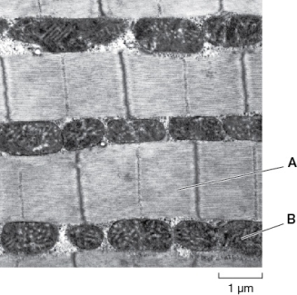
A) A = actin and myosin filaments;B = gap junctions
B) A = T tubules;B = sarcoplasm
C) A = actin and myosin filaments;B = mitochondria
D) A = T tubules;B = mitochondria
E) A = sarcoplasm;B = gap junctions
Correct Answer:

Verified
Correct Answer:
Verified
Q37: How are endoskeletal systems similar to hydrostatic
Q38: Which statement about slow-twitch and fast-twitch muscles
Q39: If a defect occurs in a developing
Q41: Smooth muscle differs from both cardiac and
Q43: Which two animals have the same type
Q44: Compared to the legs of sedentary people,the
Q45: Bricklayers occasionally need to carry stacks of
Q46: A chemical compound has been used as
Q47: Regarding skeletal,smooth,and cardiac muscle cells,which structure(s)is/are present
Q236: What is the role of the sarcoplasmic