menu-iconExamlexExamLexServices

Discover

Ask a Question
  1. All Topics
  2. Topic
    Biology
  3. Study Set
    Seeleys Anatomy Physiology Study Set 1
  4. Exam
    Exam 8: Joints and Movement
  5. Question
    -The Figure Illustrates Structures in the Right Temporomandibular Joint (Lateral
Solved

-The Figure Illustrates Structures in the Right Temporomandibular Joint (Lateral

Question 63

Question 63

Multiple Choice

  -The figure illustrates structures in the right temporomandibular joint (lateral view) .What does  D  represent? A) lateral ligament B) mandible C) zygomatic arch D) styloid process E) stylomandibular ligament
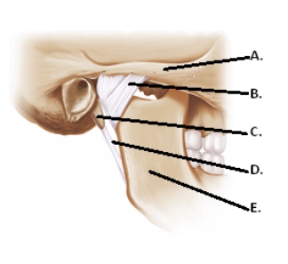
-The figure illustrates structures in the right temporomandibular joint (lateral view) .What does "D" represent?


A) lateral ligament
B) mandible
C) zygomatic arch
D) styloid process
E) stylomandibular ligament

Correct Answer:

verifed

Verified

Unlock this answer now
Get Access to more Verified Answers free of charge

Related Questions

Q10: Synovial fluid<br>A) is a double layer of

Q58: Which of the following joints is most

Q59: Which of the following joints is most

Q60: Sergio needs to reach the bowls on

Q61: A place where two or more bones

Q62: <img src="https://d2lvgg3v3hfg70.cloudfront.net/TB6158/.jpg" alt=" -The figure illustrates

Q67: <img src="https://d2lvgg3v3hfg70.cloudfront.net/TB6158/.jpg" alt=" -The figure illustrates

Q68: Which of the following does NOT occur

Q99: Hyaluronic acid<br>A) contributes to the rigidity of

Q142: The joint between the articular processes of

Examlex

ExamLex

About UsContact UsPerks CenterHomeschoolingTest Prep

Work With Us

Campus RepresentativeInfluencers

Links

FaqPricingChrome Extension

Download The App

Get App StoreGet Google Play

Policies

Privacy PolicyTerms of ServiceHonor CodeCommunity Guidelines

Scan To Download

qr-code

Copyright © (2025) ExamLex LLC.

Privacy PolicyTerms Of ServiceHonor CodeCommunity Guidelines