Multiple Choice
What anatomy and projection is demonstrated in the image below? 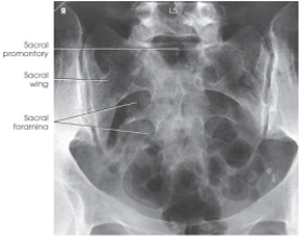
A) AP axial sacrum
B) AP oblique SI joints
C) AP coccyx
D) AP axial lumbosacral junction (Ferguson method)
Correct Answer:

Verified
Correct Answer:
Verified
Related Questions
Q127: How is the neck positioned for the
Q128: The central-ray angle for an AP axial
Q129: To demonstrate the zygapophyseal joints of the
Q130: Where is the central ray positioned for
Q131: How much is the body rotated from
Q133: Where should the superior edge of the
Q134: An abnormally increased concavity of the lumbar
Q135: Spinal nerves and blood vessels exit the
Q136: When the shoulder is immobile and cannot
Q137: The part identified on the lumbar vertebra