Multiple Choice
What projection (method) is demonstrated in the image below? 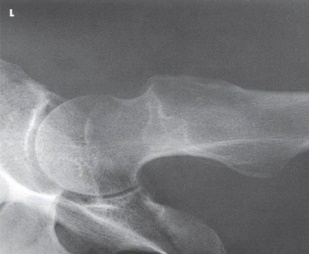
A) AP oblique (modified Cleaves)
B) Mediolateral (Lauenstein)
C) Axiolateral (Danelius-Miller)
D) AP oblique (Judet)
Correct Answer:

Verified
Correct Answer:
Verified
Related Questions
Q20: What is the recommended collimated field size
Q21: How much is the central ray angled
Q22: What positioning error is evident in the
Q23: The body is placed at what angle
Q24: The strongest bone in the body is
Q26: Which of the following will be shown
Q27: All of these comprise the hip bone,except:<br>A)
Q28: The anatomy indicated by the arrow on
Q29: Examine this AP oblique (Judet)image of the
Q30: How many degrees is the lower limb