Multiple Choice
The patient position and central-ray method demonstrated in this figure is the: 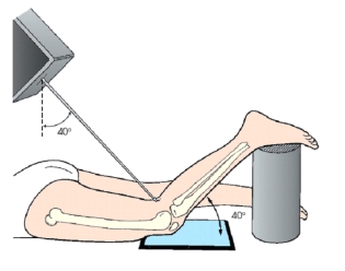
A) Holmblad (intercondylar fossa) .
B) Camp-Coventry (intercondylar fossa) .
C) Settegast (patellofemoral joint) .
D) Hughston (patellofemoral joint) .
Correct Answer:

Verified
Correct Answer:
Verified
Related Questions
Q92: The anatomy labeled with letter E in
Q93: Which of the following will clearly demonstrate
Q94: For an AP projection of the ankle,the
Q95: If a lateral projection of the femur
Q96: The projection of the patella demonstrated in
Q98: How is the patient placed for a
Q99: To better demonstrate the TMT joint spaces
Q100: The superior surface of the foot is
Q101: Where does the central ray enter the
Q102: The circular fibrocartilage disks or pads that