Multiple Choice
What projection and anatomy is demonstrated in the image below? 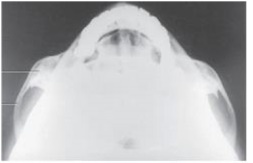
A) SMV of the TMJs
B) SMV of the zygomatic arches
C) AP axial of the TMJs
D) AP axial of the zygomatic arches
Correct Answer:

Verified
Correct Answer:
Verified
Related Questions
Q58: Which of the following projections best demonstrates
Q59: How was the central ray directed to
Q60: Which of the sinuses is developed at
Q61: The largest and most dense bone of
Q62: If the infraorbitomeatal line is placed perpendicular
Q64: The large aperture in the occipital bone,through
Q65: Letter D in the image below,used to
Q66: Which two bones are contained in the
Q67: How many bones make up the face?<br>A)
Q68: Which skull suture is found between the