Multiple Choice
Figure 10.1
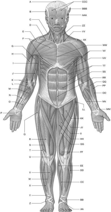
Using the above-referenced diagrammatic anterior view of the major superficial axial and appendicular muscles, identify the specified labeled item(s) in each of the following questions.
-Identify the structure(s) indicated by Label A.
A) Tendinous inscriptions
B) Epicranial aponeurosis
C) Linea alba
D) Iliotibial tract
E) Superior extensor retinaculum
Correct Answer:

Verified
Correct Answer:
Verified
Q20: The deepest lateral layer of the abdominal
Q21: The muscle that originates on the lateral
Q22: What muscular changes are involved in the
Q23: Temporalis is a muscle of facial expression,
Q24: Figure 10.4<br> <img src="https://d2lvgg3v3hfg70.cloudfront.net/TB4856/.jpg" alt="Figure 10.4
Q26: Figure 10.1<br> <img src="https://d2lvgg3v3hfg70.cloudfront.net/TB4856/.jpg" alt="Figure 10.1
Q27: Figure 10.1<br> <img src="https://d2lvgg3v3hfg70.cloudfront.net/TB4856/.jpg" alt="Figure 10.1
Q28: Figure 10.2<br> <img src="https://d2lvgg3v3hfg70.cloudfront.net/TB4856/.jpg" alt="Figure 10.2
Q29: A muscle that elevates the corner of
Q30: The deep transverse perineal muscle flexes coccygeal