Multiple Choice
Figure 10.1
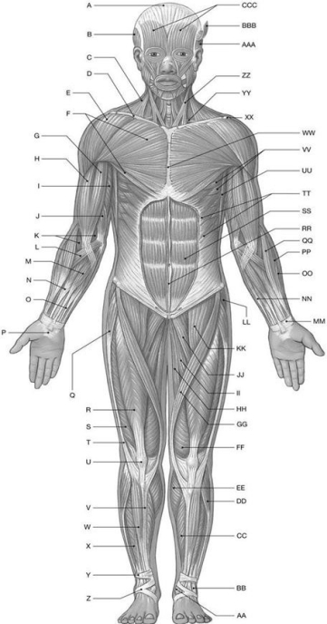
Using the above-referenced diagrammatic anterior view of the major superficial axial and appendicular muscles, identify the specified labeled item(s) in each of the following questions.
-Identify the structure(s) indicated by Label R.
A) Vastus lateralis muscle
B) Iliopsoas muscle
C) Pectineus muscle
D) Rectus femoris muscle
E) Adductor longus muscle
Correct Answer:

Verified
Correct Answer:
Verified
Q109: Which of the following can flex the
Q110: Figure 10.4<br> <img src="https://d2lvgg3v3hfg70.cloudfront.net/TB4856/.jpg" alt="Figure 10.4
Q111: The digastric muscle depresses the mandible and/or
Q112: Figure 10.4<br> <img src="https://d2lvgg3v3hfg70.cloudfront.net/TB4856/.jpg" alt="Figure 10.4
Q113: The trigeminal nerve controls which group of
Q115: Figure 10.2<br> <img src="https://d2lvgg3v3hfg70.cloudfront.net/TB4856/.jpg" alt="Figure 10.2
Q116: Which of the following belongs to the
Q117: How do the extra-ocular eye muscles differ
Q118: Why are there so few spinal flexors
Q119: The erector spinae muscle group that is