Multiple Choice
Figure 10.2
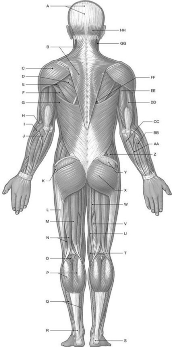
Using the above-referenced diagrammatic posterior view of the major superficial axial and appendicular muscles, identify the specified labeled item(s) in each of the following questions.
-Identify the structure(s) indicated by Label G.
A) External oblique
B) Serratus posterior
C) Latissimus dorsi
D) Rectus sheath
E) Quadratus lumborum
Correct Answer:

Verified
Correct Answer:
Verified
Q26: Figure 10.1<br> <img src="https://d2lvgg3v3hfg70.cloudfront.net/TB4856/.jpg" alt="Figure 10.1
Q27: Figure 10.1<br> <img src="https://d2lvgg3v3hfg70.cloudfront.net/TB4856/.jpg" alt="Figure 10.1
Q28: Figure 10.2<br> <img src="https://d2lvgg3v3hfg70.cloudfront.net/TB4856/.jpg" alt="Figure 10.2
Q29: A muscle that elevates the corner of
Q30: The deep transverse perineal muscle flexes coccygeal
Q32: Figure 10.4<br> <img src="https://d2lvgg3v3hfg70.cloudfront.net/TB4856/.jpg" alt="Figure 10.4
Q33: The muscle that originates on the sacrum
Q34: Figure 10.4<br> <img src="https://d2lvgg3v3hfg70.cloudfront.net/TB4856/.jpg" alt="Figure 10.4
Q35: Figure 10.4<br> <img src="https://d2lvgg3v3hfg70.cloudfront.net/TB4856/.jpg" alt="Figure 10.4
Q36: The _ muscle is divided longitudinally by