Multiple Choice
Figure 10.2
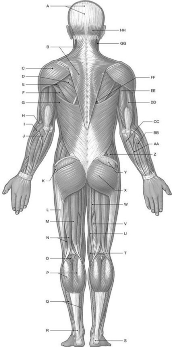
Using the above-referenced diagrammatic posterior view of the major superficial axial and appendicular muscles, identify the specified labeled item(s) in each of the following questions.
-Identify the structure(s) indicated by Label CC.
A) Flexor carpi ulnaris
B) Extensor digitorum
C) Flexor digitorum superficialis
D) Extensor carpi ulnaris
E) Flexor carpi radialis
Correct Answer:

Verified
Correct Answer:
Verified
Q116: Which of the following belongs to the
Q117: How do the extra-ocular eye muscles differ
Q118: Why are there so few spinal flexors
Q119: The erector spinae muscle group that is
Q120: Muscles of mastication most often insert into
Q122: The oblique muscle and/or rectus muscle do
Q123: Figure 10.3<br> <img src="https://d2lvgg3v3hfg70.cloudfront.net/TB4856/.jpg" alt="Figure 10.3
Q124: Figure 10.2<br> <img src="https://d2lvgg3v3hfg70.cloudfront.net/TB4856/.jpg" alt="Figure 10.2
Q125: Figure 10.2<br> <img src="https://d2lvgg3v3hfg70.cloudfront.net/TB4856/.jpg" alt="Figure 10.2
Q126: The internal intercostal muscles aid in respiration