Figure 14.1 Using the Above- Referenced Diagrams of the Superficial Anatomy and of the Superficial
Multiple Choice
Figure 14.1
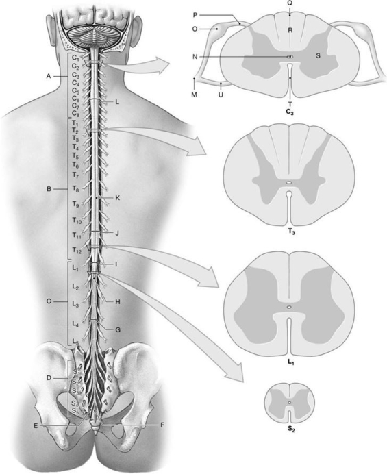
Using the above- referenced diagrams of the superficial anatomy and orientation of the adult spinal cord (posterior view) and inferior views of cross-sections through representative segments of the spinal cord, showing the arrangement of gray and white matter, identify the specified labeled structure(s) in each of the following questions.
-Identify the structure(s) indicated by Label I.
A) Cauda equina
B) Filum terminale
C) Lumbosacral enlargement
D) Conus medullaris
E) Coccygeal nerve
Correct Answer:

Verified
Correct Answer:
Verified
Q120: Figure 14.2<br> <img src="https://d2lvgg3v3hfg70.cloudfront.net/TB4856/.jpg" alt="Figure 14.2
Q121: The H-shaped mass in the center of
Q122: In step 4 of a neural reflex,
Q123: Figure 14.5<br> <img src="https://d2lvgg3v3hfg70.cloudfront.net/TB4856/.jpg" alt="Figure 14.5
Q124: The gray matter of the spinal cord
Q126: Figure 14.6<br> <img src="https://d2lvgg3v3hfg70.cloudfront.net/TB4856/.jpg" alt="Figure 14.6
Q127: Figure 14.6<br> <img src="https://d2lvgg3v3hfg70.cloudfront.net/TB4856/.jpg" alt="Figure 14.6
Q128: The first branch of each spinal nerve,
Q129: The ventral root of a spinal nerve
Q130: A normal patellar reflex indicates that spinal