Essay
Figure 4.11 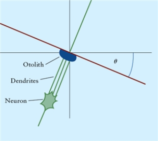
-Fig. 4.11 illustrates an otolith near the membrane surface of the utricular macula, with the head tilted sideways by angle è. Dendrites near the otolith illustrate the geometry of the otolith-membrane-dendrite interaction. Sketch the free body diagram with three forces acting on it: its weight  the normal force
  and a force parallel to the surface layer of the otolithic membrane
  Give the reasoning behind your choice of any coordinate system chosen.
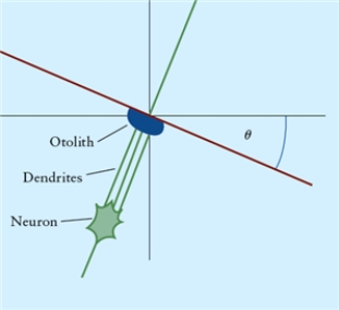
Correct Answer:

Verified
one possib...View Answer
Unlock this answer now
Get Access to more Verified Answers free of charge
Correct Answer:
Verified
View Answer
Unlock this answer now
Get Access to more Verified Answers free of charge
Q5: If you are riding on a bus
Q6: A truck has a crate in its
Q7: A sled is gliding down an incline
Q8: If an object is at rest, its
Q9: Figure 4.5 <img src="https://d2lvgg3v3hfg70.cloudfront.net/TB8418/.jpg" alt="Figure 4.5
Q11: A squid moves through the water by
Q12: Which of the following statements include all
Q13: Which one of these forces produces centripetal
Q14: If a car stays parked on an
Q15: The force of friction is parallel to