Short Answer
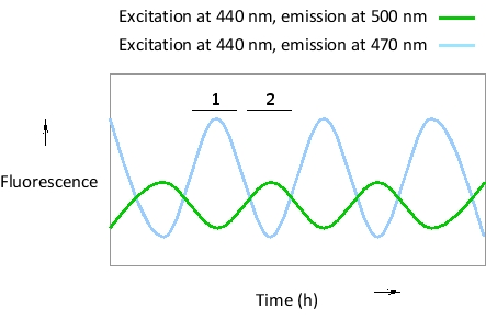
Imagine a transcription regulatory protein (X) that is known to shuttle back and forth between nucleus and cytosol in an oscillatory pattern. Protein Y is a nuclear protein that can bind to X to create a dimer that binds to DNA. You have fused protein X to green fluorescent protein (GFP) and protein Y to blue fluorescent protein (BFP), and have measured fluorescence resonance energy transfer (FRET) and non-FRET signals in the nucleus at different time points, as indicated in the following simplified plot. At which time period (1 or 2) do you think protein X is in the nucleus? BFP can be excited at 440 nm, and emits maximally at 470 nm. GFP is excited at 470 nm and emits maximally at 500 nm.
Correct Answer:

Verified
The FRET signal in the green ...View Answer
Unlock this answer now
Get Access to more Verified Answers free of charge
Correct Answer:
Verified
View Answer
Unlock this answer now
Get Access to more Verified Answers free of charge
Q16: Indicate true (T) and false (F) statements
Q17: Single-molecule detection by fluorescence microscopy is limited
Q18: Two approaches have been devised to deal
Q19: The following schematic diagram shows the path
Q20: Indicate whether you would use a fluorescent
Q22: In which of the following microscopy techniques
Q23: If an average globular protein was of
Q24: The light used to excite a fluorescent
Q25: Tubulin labeled with caged fluorescein can be
Q26: Two segments (S1 and S2) in a