Multiple Choice
What is the projection (method) demonstrated in the image below used to evaluate the cranium?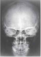
A) Parietoacanthial (Waters)
B) PA axial (Caldwell)
C) AP axial (Towne)
D) PA
Correct Answer:

Verified
Correct Answer:
Verified
Related Questions
Q21: Which of the following is true regarding
Q23: The suture located between the occipital bone
Q24: The opening into the apex of the
Q25: The part of the sphenoid bone identified
Q27: What structure is labeled as letter B
Q30: What type of joint is the TMJ?<br>A)
Q32: The parietoacanthial projection (Waters method)of the sinuses
Q51: Which line should be placed parallel to
Q78: The posterior half of the base of
Q103: What is the central-ray angulation for the