Multiple Choice
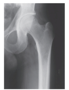
-Refer to the figure. What can be said about the image of cortical margins of the femur?
A) The thickness of the cortical image is normal
B) The absence of cortical image at the femoral neck indicates a demineralized state
C) The thickness of the cortical margins is pronounced, indicating a bone deposition disease
D) The pencil-thin cortical margins on the shaft indicate osteoporosis
Correct Answer:

Verified
Correct Answer:
Verified
Q1: <img src="https://d2lvgg3v3hfg70.cloudfront.net/TB11028/.jpg" alt=" -You recognize the
Q2: <img src="https://d2lvgg3v3hfg70.cloudfront.net/TB11028/.jpg" alt=" -Refer to the
Q3: <img src="https://d2lvgg3v3hfg70.cloudfront.net/TB11028/.jpg" alt=" -Refer to the
Q4: <img src="https://d2lvgg3v3hfg70.cloudfront.net/TB11028/.jpg" alt=" -Refer to the
Q6: Axial migration of the femoral head is
Q7: Which of the following is true regarding
Q8: <img src="https://d2lvgg3v3hfg70.cloudfront.net/TB11028/.jpg" alt=" -Refer to the
Q9: Which of the following pathological conditions is
Q10: <img src="https://d2lvgg3v3hfg70.cloudfront.net/TB11028/.jpg" alt=" -Refer to the