Multiple Choice
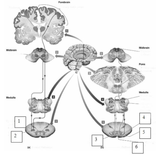
-The pathways illustrated in the left side of this figure (numbers 1 and 2) are known as the
A) medial lemniscus.
B) spinothalamic pathways.
C) lateral pathways.
D) ventromedial pathways.
Correct Answer:

Verified
Correct Answer:
Verified
Related Questions
Q19: The lateral pathway originates in the _
Q39: Exposure to environmental toxins may lead to<br>A)
Q40: A single motor unit usually contains a
Q55: One of the actions of the basal
Q66: Describe the physical symptoms of myasthenia gravis.
Q70: Alpha motor neurons are <br>A)small and unmyelinated. <br>B)small and
Q71: Elizabeth's grandmother recently underwent a hip replacement
Q72: Many toxins responsible for motor disorders interact
Q73: Rigor mortis,or the muscle stiffness present after
Q121: _ is a protein that makes up