Multiple Choice
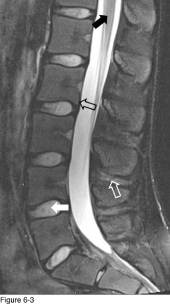
-Identify the diagnostic image in Figure 6-3.
A) T2 sagittal lumbar MRI
B) T1 sagittal lumbar MRI
C) Bone window sagittal reconstruct CT
D) Soft-tissue window sagittal reconstruct CT
Correct Answer:

Verified
Correct Answer:
Verified
Q4: <img src="https://d2lvgg3v3hfg70.cloudfront.net/TB4823/.jpg" alt=" -The structure indicated
Q5: <img src="https://d2lvgg3v3hfg70.cloudfront.net/TB4823/.jpg" alt=" -The structure marked
Q6: <img src="https://d2lvgg3v3hfg70.cloudfront.net/TB4823/.jpg" alt=" -The structure indicated
Q7: <img src="https://d2lvgg3v3hfg70.cloudfront.net/TB4823/.jpg" alt=" -The structure indicated
Q8: <img src="https://d2lvgg3v3hfg70.cloudfront.net/TB4823/.jpg" alt=" -The structure indicated
Q10: <img src="https://d2lvgg3v3hfg70.cloudfront.net/TB4823/.jpg" alt=" -What type of
Q11: <img src="https://d2lvgg3v3hfg70.cloudfront.net/TB4823/.jpg" alt=" -The structure indicated
Q12: <img src="https://d2lvgg3v3hfg70.cloudfront.net/TB4823/.jpg" alt=" -Figure 6-1 is
Q13: <img src="https://d2lvgg3v3hfg70.cloudfront.net/TB4823/.jpg" alt=" -The structure indicated
Q14: <img src="https://d2lvgg3v3hfg70.cloudfront.net/TB4823/.jpg" alt=" -What structure is