Essay
West Nile virus (WNV) is a human pathogen that is transmitted by mosquitoes, which inject the virus into the host while they are feeding. To study the immune response to WNV, mice can be infected by injecting 100 PFU of virus into the footpad. This generates a robust immune response 7 days post-infection in the popliteal lymph node (LN), located behind the knee, as indicated in Figure. 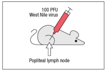
Popliteal LN cells are isolated from the mouse shown above at day 7 post-infection, and the T cells are stimulated in vitro with the WNV peptide P33 plus dendritic cells isolated from a non-infected mouse of the same strain. Five hours later, the CD8 T cells in the culture are analyzed for their ability to produce the cytokine IFN- , and the numbers of IFN- -producing CD8 T cells are quantified. As a control, T cells are also stimulated with an irrelevant non-viral peptide (ova) plus dendritic cells. The results are shown in Figure. 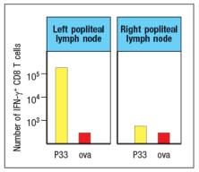
a) Why is the T cell response different between the two lymph node populations?
To identify the important antigen-presenting cell required to activate WNV-specific CD8 T cells in the popliteal LN, wild-type mice and Batf3-/- mice are each immunized with 100 PFU of WNV in the left footpad, as above. Previous studies have indicated that Batf3-/- mice lack one particular subset of conventional dendritic cells, known as CD8 + dendritic cells (DC), but otherwise appear to have normal numbers and subsets of all other immune cell populations (e.g., T cells, B cells, macrophages, etc.). The results of this experiment are shown in Figure
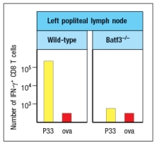
b) Name two possible functions of CD8 + dendritic cells that could account for the results seen in the Batf3-/- mice immunized with WNV.
To distinguish between the possible functions of CD8 + dendritic cells, another set of wild-type and Batf3-/- mice are immunized with 100 PFU of WNV in the left footpad. At day 7 post-infection, the left popliteal LN are isolated from the mice, and the CD4 T cells in these LN populations are tested for responses to a WNV peptide bound to MHC class II (peptide Y). As a negative control, an MHC class II-binding peptide from ova is used. These results are shown in Figure .
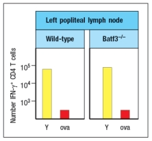
c) Do these results rule in/out either of your proposed functions of CD8 + dendritic cells indicated in answer to part (b)? Why or why not?
Correct Answer:

Verified
a) The immunization with the WNV was in ...View Answer
Unlock this answer now
Get Access to more Verified Answers free of charge
Correct Answer:
Verified
View Answer
Unlock this answer now
Get Access to more Verified Answers free of charge
Q4: Wiskott-Aldrich syndrome is an immunodeficiency disease that
Q5: Inbred strains of mice often generate highly
Q6: Eosinophils are a subset of granulocytes
Q7: The compound LE135 is an inhibitor
Q8: Many virus infections induce robust production
Q10: A mouse is infected with staphylococcal bacteria
Q11: In a lymph node, nT<sub>reg</sub> cells are
Q12: When T cells are activated by recognizing
Q13: Each subset of effector T cells produces
Q14: An immunodeficient mouse strain is identified,