Deck 4: Microscopy
Full screen (f)
Question
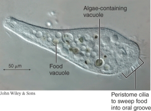
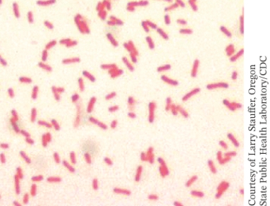
Using the information from the image below, determine the magnification of the microbe.
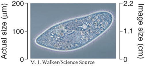
A) 200X
B) 2.2X
C) 91X
D) 110X
A) 200X
B) 2.2X
C) 91X
D) 110X
Question
Question
Question
Question
Question
Question
Question
Question
Question
Question
Question
The image below represents a(n): ________.
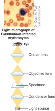
A) Dark field microscope
B) Bright field microscope
C) Fluorescence microscope
D) Phase contrast microscope
A) Dark field microscope
B) Bright field microscope
C) Fluorescence microscope
D) Phase contrast microscope
Question
Question
Question
What type of microscope was used to produce the image below?
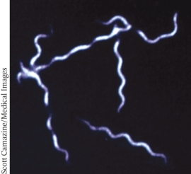
A) Dark field microscope
B) Bright field microscope
C) Fluorescence microscope
D) Phase contrast microscope
A) Dark field microscope
B) Bright field microscope
C) Fluorescence microscope
D) Phase contrast microscope
Question
Question
Question
What type of microscope is shown below?
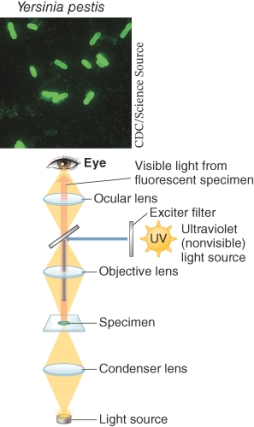
A) Bright field microscope
B) Transmission electron microscope
C) Dark field microscope
D) Fluorescence microscope
A) Bright field microscope
B) Transmission electron microscope
C) Dark field microscope
D) Fluorescence microscope
Question
Question
Question
What type of microscope was used to produce the image below?
A) Differential interference contrast microscopy
B) Bright field microscope
C) Fluorescence microscope
D) Confocal microscope
A) Differential interference contrast microscopy
B) Bright field microscope
C) Fluorescence microscope
D) Confocal microscope
Question
Question
Question
Question
Question
What type of microscope is shown below?
A) Bright field microscope
B) Transmission electron microscope
C) Fluorescence microscope
D) Dark field microscope
A) Bright field microscope
B) Transmission electron microscope
C) Fluorescence microscope
D) Dark field microscope
Question
What type of microscope is shown below?
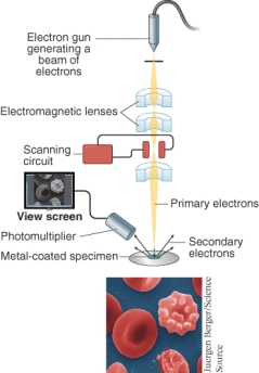
A) Dark field microscope
B) Transmission electron microscope
C) Scanning electron microscope
D) Fluorescence microscope
A) Dark field microscope
B) Transmission electron microscope
C) Scanning electron microscope
D) Fluorescence microscope
Question
Question
Question
Question
Question
Question
Question
Question
Question
Question
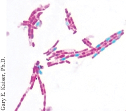
The bright field light micrograph below is an image of: ________.
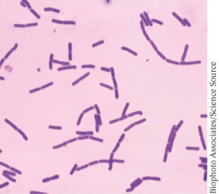
A) gram positive bacterial cells
B) gram negative bacterial cells
C) acid fast bacterial cells
D) bacterial cells with endospores
A) gram positive bacterial cells
B) gram negative bacterial cells
C) acid fast bacterial cells
D) bacterial cells with endospores
Question
The bright field light micrograph below is an image of: ________.
A) gram positive bacterial cells
B) gram negative bacterial cells
C) acid fast bacterial cells
D) bacterial cells with endospores
A) gram positive bacterial cells
B) gram negative bacterial cells
C) acid fast bacterial cells
D) bacterial cells with endospores
Question
A 20-year-old male presented at the emergency room with intense burning during urination. A copious amount of pus was being discharged from his penis. A micrograph of a gram stain of the pus is shown below. The man is infected with ________.
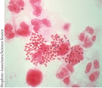
A) gram negative bacteria that have infected leukocytes
B) gram positive bacteria that have infected leukocytes
C) gram positive bacteria and gram positive eukaryotic pathogens
D) gram negative bacteria and gram negative eukaryotic pathogens
A) gram negative bacteria that have infected leukocytes
B) gram positive bacteria that have infected leukocytes
C) gram positive bacteria and gram positive eukaryotic pathogens
D) gram negative bacteria and gram negative eukaryotic pathogens
Question
Question
Question
Question
Question
Question
Question
Question
Question
Question
The light micrograph below is an example of: ________.
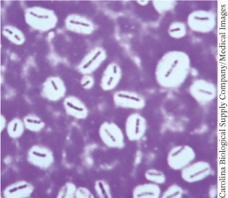
A) A capsule stain
B) An acid fast stain
C) An endospore stain
D) Dark field microscopy
A) A capsule stain
B) An acid fast stain
C) An endospore stain
D) Dark field microscopy
Question
The light micrograph below is an example of: ________.
A) A capsule stain
B) An acid fast stain
C) An endospore stain
D) A Gram stain
A) A capsule stain
B) An acid fast stain
C) An endospore stain
D) A Gram stain
Question
Question
Question

Unlock Deck
Sign up to unlock the cards in this deck!
Unlock Deck
Unlock Deck
1/53
Play
Full screen (f)
Deck 4: Microscopy
1
Using the information from the image below, determine the magnification of the microbe.
A) 200X
B) 2.2X
C) 91X
D) 110X
A) 200X
B) 2.2X
C) 91X
D) 110X
D
2
In the history of microscopy, who is considered the "Father of Microbiology"?
A) Hans Jansen
B) Zacharias Jansen
C) Antonie van Leeuwenhoek
D) Ernst Ruska
A) Hans Jansen
B) Zacharias Jansen
C) Antonie van Leeuwenhoek
D) Ernst Ruska
C
3
What is the definition of the term magnification?
A) Ratio of the size of the bacterium to the size of the object.
B) Ratio of the size of the protozoan to the size of the objective.
C) Ratio of the size of the image to the size of the object.
D) Ratio of the size of the objective to the size of the microorganism.
A) Ratio of the size of the bacterium to the size of the object.
B) Ratio of the size of the protozoan to the size of the objective.
C) Ratio of the size of the image to the size of the object.
D) Ratio of the size of the objective to the size of the microorganism.
C
4
The iris diaphragm ________.
A) focuses light on specimen
B) separately magnify specimen
C) holds the specimen in place
D) controls light intensity via adjustable opening
A) focuses light on specimen
B) separately magnify specimen
C) holds the specimen in place
D) controls light intensity via adjustable opening
Unlock Deck
Unlock for access to all 53 flashcards in this deck.
Unlock Deck
k this deck
5
The condenser ________.
A) focuses light on specimen
B) separately magnify specimen
C) holds the specimen in place
D) controls light intensity via adjustable opening
A) focuses light on specimen
B) separately magnify specimen
C) holds the specimen in place
D) controls light intensity via adjustable opening
Unlock Deck
Unlock for access to all 53 flashcards in this deck.
Unlock Deck
k this deck
6
The ________ holds the specimen in place on a light microscope.
A) Iris diaphragm
B) Condenser lens
C) Objective and Ocular lenses
D) Stage
A) Iris diaphragm
B) Condenser lens
C) Objective and Ocular lenses
D) Stage
Unlock Deck
Unlock for access to all 53 flashcards in this deck.
Unlock Deck
k this deck
7
The________ magnify the specimen on a light microscope.
A) Iris diaphragm
B) Condenser lens
C) Objective and Ocular lenses
D) Stage
A) Iris diaphragm
B) Condenser lens
C) Objective and Ocular lenses
D) Stage
Unlock Deck
Unlock for access to all 53 flashcards in this deck.
Unlock Deck
k this deck
8
What is the correct order of microscope parts from the base to the ocular?
A) Base, illuminator, condenser lens, iris diaphragm, stage, objective lens, ocular lens
B) Base, condenser lens, illuminator, iris diaphragm, objective lens, stage, ocular lens
C) Base, iris diaphragm, stage, illuminator, condenser lens, objective lens, ocular lens
D) Base, stage, condenser lens, illuminator, objective lens, stage, iris diaphragm, ocular lens
Unlock Deck
Unlock for access to all 53 flashcards in this deck.
Unlock Deck
k this deck
9
What two features of a microscope are essential for a microbiologist?
A) Magnification and resolution
B) Magnification and oil immersion
C) Resolution and oil immersion
D) Objectives and oculars
A) Magnification and resolution
B) Magnification and oil immersion
C) Resolution and oil immersion
D) Objectives and oculars
Unlock Deck
Unlock for access to all 53 flashcards in this deck.
Unlock Deck
k this deck
10
Which of the following best defines the term "resolution"?
A) The degree to which a microscope shows two images very far apart
B) The degree to which a microscope shows two or more cells as being distinct
C) The degree to which a microscope shows two or more cells as being close together
D) The degree to which a microscope shows two very close objects as being separate and apart
Unlock Deck
Unlock for access to all 53 flashcards in this deck.
Unlock Deck
k this deck
11
What is the effect of using immersion oil plus oil immersion objective? (Select all that apply)
A) Resolving power is increased because immersion oil has same refractive index as glass.
B) As light passes through the glass of the cover slip, oil and the glass of the oil immersion lens, there is no light lost as a result of differences in refractive index.
C) Increases both magnification and resolving power of microscope.
D) Magnification increases from 1000X to 4500X.
Unlock Deck
Unlock for access to all 53 flashcards in this deck.
Unlock Deck
k this deck
12
The image below represents a(n): ________.
A) Dark field microscope
B) Bright field microscope
C) Fluorescence microscope
D) Phase contrast microscope
A) Dark field microscope
B) Bright field microscope
C) Fluorescence microscope
D) Phase contrast microscope
Unlock Deck
Unlock for access to all 53 flashcards in this deck.
Unlock Deck
k this deck
13
A bright field microscope ________. (Select all that apply)
A) produces an image showing fluorescent portions of specimen
B) is the simplest, most common type of microscope
C) produces a bright image against dark background
D) produces the image of a stained specimen is visualized against a bright background
A) produces an image showing fluorescent portions of specimen
B) is the simplest, most common type of microscope
C) produces a bright image against dark background
D) produces the image of a stained specimen is visualized against a bright background
Unlock Deck
Unlock for access to all 53 flashcards in this deck.
Unlock Deck
k this deck
14
What are the disadvantages of Bright-field microscopy? (Select all that apply)
A) Staining is required to make specimens visible
B) The limit for magnification is 1000X
C) Images of very small specimens show very little detail, just the overall organism structure
D) It is the simplest, most common type of microscope
A) Staining is required to make specimens visible
B) The limit for magnification is 1000X
C) Images of very small specimens show very little detail, just the overall organism structure
D) It is the simplest, most common type of microscope
Unlock Deck
Unlock for access to all 53 flashcards in this deck.
Unlock Deck
k this deck
15
What type of microscope was used to produce the image below?
A) Dark field microscope
B) Bright field microscope
C) Fluorescence microscope
D) Phase contrast microscope
A) Dark field microscope
B) Bright field microscope
C) Fluorescence microscope
D) Phase contrast microscope
Unlock Deck
Unlock for access to all 53 flashcards in this deck.
Unlock Deck
k this deck
16
A dark field microscope ________.
A) produces an image showing fluorescent portions of specimen
B) is the simplest, most common type of microscope
C) produces a bright image against dark background
D) produces the image of a stained specimen is visualized against a bright background
A) produces an image showing fluorescent portions of specimen
B) is the simplest, most common type of microscope
C) produces a bright image against dark background
D) produces the image of a stained specimen is visualized against a bright background
Unlock Deck
Unlock for access to all 53 flashcards in this deck.
Unlock Deck
k this deck
17
What are the advantages of Dark-field microscopy? (Select all that apply)
A) It may be used for living microbes
B) It may be used for hard-to-stain microbes
C) It highlights only surfaces of microbes
D) It can be used to visualize very thin specimens
A) It may be used for living microbes
B) It may be used for hard-to-stain microbes
C) It highlights only surfaces of microbes
D) It can be used to visualize very thin specimens
Unlock Deck
Unlock for access to all 53 flashcards in this deck.
Unlock Deck
k this deck
18
What type of microscope is shown below?
A) Bright field microscope
B) Transmission electron microscope
C) Dark field microscope
D) Fluorescence microscope
A) Bright field microscope
B) Transmission electron microscope
C) Dark field microscope
D) Fluorescence microscope
Unlock Deck
Unlock for access to all 53 flashcards in this deck.
Unlock Deck
k this deck
19
What dyes are used most often with fluorescence microscopy?
A) Malachite green and carbol fuchsin
B) Fluorescein and rhodamine
C) Fluorescein and crystal violet
D) Crystal violet and safranin
A) Malachite green and carbol fuchsin
B) Fluorescein and rhodamine
C) Fluorescein and crystal violet
D) Crystal violet and safranin
Unlock Deck
Unlock for access to all 53 flashcards in this deck.
Unlock Deck
k this deck
20
Which of the following statements is TRUE concerning Direct Fluorescent Antibody (DFA) Assay use in microscopy? (Select all that apply)
A) Monoclonal antibodies bind to dyes and are then bound to pathogens
B) Antibody-dye conjugates glow under uv light and show presence of pathogen
C) Fluorescein and rhodamine are dyes most commonly used as they glow green and red
D) It is the simplest, most common type of microscope
A) Monoclonal antibodies bind to dyes and are then bound to pathogens
B) Antibody-dye conjugates glow under uv light and show presence of pathogen
C) Fluorescein and rhodamine are dyes most commonly used as they glow green and red
D) It is the simplest, most common type of microscope
Unlock Deck
Unlock for access to all 53 flashcards in this deck.
Unlock Deck
k this deck
21
What type of microscope was used to produce the image below?
A) Differential interference contrast microscopy
B) Bright field microscope
C) Fluorescence microscope
D) Confocal microscope
A) Differential interference contrast microscopy
B) Bright field microscope
C) Fluorescence microscope
D) Confocal microscope
Unlock Deck
Unlock for access to all 53 flashcards in this deck.
Unlock Deck
k this deck
22
Confocal Microscopy: ________.
A) Uses an electrically charged nanoprobe to scan the surface of a metal-coated specimen
B) Produces a three-dimensional image by assembling scanned regions of a fluorescent specimen
C) Displays unstained cellular structures that are visualized using differences in refractive indices
D) An ultrathin specimen is penetrated by an electron beam to show very good magnification and detailed resolution
Unlock Deck
Unlock for access to all 53 flashcards in this deck.
Unlock Deck
k this deck
23
Phase Contrast Microscopy: ________.
A) Uses an electrically charged nanoprobe to scan the surface of a metal-coated specimen
B) Produces a three-dimensional image by assembling scanned regions of a fluorescent specimen
C) Displays unstained cellular structures that are visualized using differences in refractive indices
D) An ultrathin specimen is penetrated by an electron beam to show very good magnification and detailed resolution
Unlock Deck
Unlock for access to all 53 flashcards in this deck.
Unlock Deck
k this deck
24
What are the major characteristics of phase contrast microscopy? (Select all that apply)
A) The true structure of living specimens may be viewed without the potential distortion of staining.
B) It converts differences in refractive indices to differences in color via use of prisms.
C) Special filters convert differences in light that are out of phase with waves through background into differences in light intensity.
D) It increases the resolution limit of a confocal microscope.
Unlock Deck
Unlock for access to all 53 flashcards in this deck.
Unlock Deck
k this deck
25
Differential Interference Contrast Microscopy (DIC) makes use of these features to enable microbiologists to view a living specimen: ________.
A) changes in light waves as they pass through the living specimen are out of phase with each other
B) utilizes a bright-field microscope with filters
C) Three-dimensional color images generated
D) Magnification is higher than that of a bright-field microscope
A) changes in light waves as they pass through the living specimen are out of phase with each other
B) utilizes a bright-field microscope with filters
C) Three-dimensional color images generated
D) Magnification is higher than that of a bright-field microscope
Unlock Deck
Unlock for access to all 53 flashcards in this deck.
Unlock Deck
k this deck
26
What type of microscope is shown below?
A) Bright field microscope
B) Transmission electron microscope
C) Fluorescence microscope
D) Dark field microscope
A) Bright field microscope
B) Transmission electron microscope
C) Fluorescence microscope
D) Dark field microscope
Unlock Deck
Unlock for access to all 53 flashcards in this deck.
Unlock Deck
k this deck
27
What type of microscope is shown below?
A) Dark field microscope
B) Transmission electron microscope
C) Scanning electron microscope
D) Fluorescence microscope
A) Dark field microscope
B) Transmission electron microscope
C) Scanning electron microscope
D) Fluorescence microscope
Unlock Deck
Unlock for access to all 53 flashcards in this deck.
Unlock Deck
k this deck
28
An ultrathin specimen is penetrated by transmission of an electron beam to produce and image with high magnification and detailed resolution.
A) Confocal Microscopy
B) Phase Contrast Microscopy
C) Transmission Electron Microscopy
D) Scanning Electron Microscopy
A) Confocal Microscopy
B) Phase Contrast Microscopy
C) Transmission Electron Microscopy
D) Scanning Electron Microscopy
Unlock Deck
Unlock for access to all 53 flashcards in this deck.
Unlock Deck
k this deck
29
Electrons are scattered from the surface of a metal-coated specimen to generate a three-dimensional image with high magnification and detailed resolution.
A) Confocal Microscopy
B) Phase Contrast Microscopy
C) Transmission Electron Microscopy
D) Scanning Electron Microscopy
A) Confocal Microscopy
B) Phase Contrast Microscopy
C) Transmission Electron Microscopy
D) Scanning Electron Microscopy
Unlock Deck
Unlock for access to all 53 flashcards in this deck.
Unlock Deck
k this deck
30
What of the following is a correct statement about the major differences between electron and light microscopes? (Select all that apply)
A) Electron microscopes use beams of electrons and electromagnetic lenses while light microscopes use visible light waves and glass lenses.
B) Electron microscopes can show detailed images at the level of the cellular organelles while light microscopes cannot do this.
C) Electron microscopes have a lower resolving power that light microscopes because an electron bean has a longer wavelength that visible light.
D) Electron microscopes produces images on a photographic plate while light microscopes produce images through the ocular lens.
Unlock Deck
Unlock for access to all 53 flashcards in this deck.
Unlock Deck
k this deck
31
What is the major difference between TEM and SEM?
A) TEM uses light waves and electrons while SEM uses only electron beams.
B) TEM uses electromagnetic condenser lens as well as an electromagnetic objective lens while SEM uses only 2 electromagnetic lenses.
C) TEM uses a projector lens while SEM uses a photomultiplier.
D) TEM projects a 2-dimensional cross section of a microbe while SEM projects a 3-dimensional image with surface details.
Unlock Deck
Unlock for access to all 53 flashcards in this deck.
Unlock Deck
k this deck
32
Which of the following is a major characteristic of super-resolution microscopy? (Select all that apply)
A) It increases the resolution of transmission electron microscopy
B) It increases the resolution limit of a confocal microscope
C) It is able to produce an image in very low light
D) It can visualize living and dead cells
A) It increases the resolution of transmission electron microscopy
B) It increases the resolution limit of a confocal microscope
C) It is able to produce an image in very low light
D) It can visualize living and dead cells
Unlock Deck
Unlock for access to all 53 flashcards in this deck.
Unlock Deck
k this deck
33
The very complex microscopes used in research are classified according to ________.
A) how large and complex they are
B) how they generate a magnified image
C) type of stain required
D) whether they make use of probes
A) how large and complex they are
B) how they generate a magnified image
C) type of stain required
D) whether they make use of probes
Unlock Deck
Unlock for access to all 53 flashcards in this deck.
Unlock Deck
k this deck
34
What is the incorrect match below?
A) Light microscope/visible light and glass lenses.
B) Electron microscope/electron beam and electromagnetic lenses.
C) Nanoprobe microscope/ fine-tipped probes are used to penetrate the surface of a specimen to magnify internal structures.
D) Confocal microscope/illuminates one focal plane at a time which is then reassembled into a three-dimensional image.
Unlock Deck
Unlock for access to all 53 flashcards in this deck.
Unlock Deck
k this deck
35
Which of the following statements about Scanning Tunneling Microscopy is correct? (Select all that apply)
A) It utilizes an electrically charged nanoprobe to scan surface of a metal-coated specimen.
B) It measures changes in voltage detected by the nanoprobe.
C) The voltage changes detected by the probe are converted to an image.
D) The resolution is not good enough to see anything at the atomic level.
A) It utilizes an electrically charged nanoprobe to scan surface of a metal-coated specimen.
B) It measures changes in voltage detected by the nanoprobe.
C) The voltage changes detected by the probe are converted to an image.
D) The resolution is not good enough to see anything at the atomic level.
Unlock Deck
Unlock for access to all 53 flashcards in this deck.
Unlock Deck
k this deck
36
Which of the following statements about Atomic Force Microscopy is true? (Select all that apply)
A) A fine-tipped probe is dragged across surface of molecules like proteins.
B) The movements of probe are amplified to produce an image.
C) The heights of a structure can be delineated relative to the background.
D) The voltage changes detected by the probe are converted to an image.
A) A fine-tipped probe is dragged across surface of molecules like proteins.
B) The movements of probe are amplified to produce an image.
C) The heights of a structure can be delineated relative to the background.
D) The voltage changes detected by the probe are converted to an image.
Unlock Deck
Unlock for access to all 53 flashcards in this deck.
Unlock Deck
k this deck
37
The bright field light micrograph below is an image of: ________.
A) gram positive bacterial cells
B) gram negative bacterial cells
C) acid fast bacterial cells
D) bacterial cells with endospores
A) gram positive bacterial cells
B) gram negative bacterial cells
C) acid fast bacterial cells
D) bacterial cells with endospores
Unlock Deck
Unlock for access to all 53 flashcards in this deck.
Unlock Deck
k this deck
38
The bright field light micrograph below is an image of: ________.
A) gram positive bacterial cells
B) gram negative bacterial cells
C) acid fast bacterial cells
D) bacterial cells with endospores
A) gram positive bacterial cells
B) gram negative bacterial cells
C) acid fast bacterial cells
D) bacterial cells with endospores
Unlock Deck
Unlock for access to all 53 flashcards in this deck.
Unlock Deck
k this deck
39
A 20-year-old male presented at the emergency room with intense burning during urination. A copious amount of pus was being discharged from his penis. A micrograph of a gram stain of the pus is shown below. The man is infected with ________.
A) gram negative bacteria that have infected leukocytes
B) gram positive bacteria that have infected leukocytes
C) gram positive bacteria and gram positive eukaryotic pathogens
D) gram negative bacteria and gram negative eukaryotic pathogens
A) gram negative bacteria that have infected leukocytes
B) gram positive bacteria that have infected leukocytes
C) gram positive bacteria and gram positive eukaryotic pathogens
D) gram negative bacteria and gram negative eukaryotic pathogens
Unlock Deck
Unlock for access to all 53 flashcards in this deck.
Unlock Deck
k this deck
40
What is the correct order of steps in smear preparation?
1) Apply suspension of cells on slide
2) spread over surface of slide
3) heat fix or chemical fix specimen on slide
4) air dry
A) 1, 2, 4, 3
B) 1, 2, 3, 4
C) 1, 3, 2, 4
D) 3, 2, 1, 4
1) Apply suspension of cells on slide
2) spread over surface of slide
3) heat fix or chemical fix specimen on slide
4) air dry
A) 1, 2, 4, 3
B) 1, 2, 3, 4
C) 1, 3, 2, 4
D) 3, 2, 1, 4
Unlock Deck
Unlock for access to all 53 flashcards in this deck.
Unlock Deck
k this deck
41
Simple Stains have this characteristic: ________. (Select all that apply)
A) Only one dye is used
B) Staining will show size, shape and arrangement of cells
C) They use a decolorizer
D) they use a secondary stain
A) Only one dye is used
B) Staining will show size, shape and arrangement of cells
C) They use a decolorizer
D) they use a secondary stain
Unlock Deck
Unlock for access to all 53 flashcards in this deck.
Unlock Deck
k this deck
42
Positive Staining technique: ________.
A) shows clear cells against a darkly stained background
B) uses stains that are acidic such as nigrosine and Indian ink
C) uses stains that have a negative charge that is repelled by the negative charge of cell surface carbohydrates and glycoproteins
D) uses basic dyes such as Methylene Blue and Crystal Violet
Unlock Deck
Unlock for access to all 53 flashcards in this deck.
Unlock Deck
k this deck
43
Among the characteristics of Positive Staining are: ________. (Select all that apply)
A) cells are colored against a clear background
B) The positive charge of stain is attracted to negatively charges on DNA and surface proteins
C) examples include nigrosin and India ink
D) Examples include methylene blue and crystal violet
A) cells are colored against a clear background
B) The positive charge of stain is attracted to negatively charges on DNA and surface proteins
C) examples include nigrosin and India ink
D) Examples include methylene blue and crystal violet
Unlock Deck
Unlock for access to all 53 flashcards in this deck.
Unlock Deck
k this deck
44
Negative staining: ________. (Select all that apply)
A) shows clear cells against darkly stained background
B) uses stains that are acidic and have a negative charge
C) works because the negative charge is repelled by negative charge of carbs and glycoproteins on bacterial surfaces
D) shows stained cells against a clear background
A) shows clear cells against darkly stained background
B) uses stains that are acidic and have a negative charge
C) works because the negative charge is repelled by negative charge of carbs and glycoproteins on bacterial surfaces
D) shows stained cells against a clear background
Unlock Deck
Unlock for access to all 53 flashcards in this deck.
Unlock Deck
k this deck
45
Differential staining requires: ________. (Select all that apply)
A) preparation of a smear
B) application of 2 or more different positive stains
C) decolorizing chemical
D) preparation of a hanging drop
A) preparation of a smear
B) application of 2 or more different positive stains
C) decolorizing chemical
D) preparation of a hanging drop
Unlock Deck
Unlock for access to all 53 flashcards in this deck.
Unlock Deck
k this deck
46
What is the primary stain used in the Gram stain?
A) Crystal violet
B) Safranin
C) Malachite Green
D) Methylene Blue
A) Crystal violet
B) Safranin
C) Malachite Green
D) Methylene Blue
Unlock Deck
Unlock for access to all 53 flashcards in this deck.
Unlock Deck
k this deck
47
What is the primary stain used in the Acid-Fast stain?
A) Crystal violet
B) Carbol fuchsin
C) Malachite Green
D) Methylene Blue
A) Crystal violet
B) Carbol fuchsin
C) Malachite Green
D) Methylene Blue
Unlock Deck
Unlock for access to all 53 flashcards in this deck.
Unlock Deck
k this deck
48
What is the counter or secondary stain used in the Acid-Fast stain?
A) Crystal violet
B) Safranin
C) Carbol fuchsin
D) Methylene Blue
A) Crystal violet
B) Safranin
C) Carbol fuchsin
D) Methylene Blue
Unlock Deck
Unlock for access to all 53 flashcards in this deck.
Unlock Deck
k this deck
49
The light micrograph below is an example of: ________.
A) A capsule stain
B) An acid fast stain
C) An endospore stain
D) Dark field microscopy
A) A capsule stain
B) An acid fast stain
C) An endospore stain
D) Dark field microscopy
Unlock Deck
Unlock for access to all 53 flashcards in this deck.
Unlock Deck
k this deck
50
The light micrograph below is an example of: ________.
A) A capsule stain
B) An acid fast stain
C) An endospore stain
D) A Gram stain
A) A capsule stain
B) An acid fast stain
C) An endospore stain
D) A Gram stain
Unlock Deck
Unlock for access to all 53 flashcards in this deck.
Unlock Deck
k this deck
51
Capsule staining shows ________ capsules surrounding ________ cells against a ________ background.
A) clear; colored; colored
B) colored; clear; colored
C) colored; colored; clear
D) clear; colored; clear
A) clear; colored; colored
B) colored; clear; colored
C) colored; colored; clear
D) clear; colored; clear
Unlock Deck
Unlock for access to all 53 flashcards in this deck.
Unlock Deck
k this deck
52
Flagella staining requires ________ to make the flagella more visible.
A) dye only in a single layer
B) dye plus mordent in a single layer of dye
C) mordent only in a single layer
D) dye plus mordant in multiple layers
A) dye only in a single layer
B) dye plus mordent in a single layer of dye
C) mordent only in a single layer
D) dye plus mordant in multiple layers
Unlock Deck
Unlock for access to all 53 flashcards in this deck.
Unlock Deck
k this deck
53
What is the primary stain used in the endospore stain?
A) Safranin
B) Carbol fuchsin
C) Malachite Green
D) Methylene Blue
A) Safranin
B) Carbol fuchsin
C) Malachite Green
D) Methylene Blue
Unlock Deck
Unlock for access to all 53 flashcards in this deck.
Unlock Deck
k this deck
Unlock Deck
Unlock for access to all 53 flashcards in this deck.


