Exam 6: Anatomy Layering and Sectional Anatomy
Exam 1: Before and After the Ultrasound Examination30 Questions
Exam 2: Ultrasound Instrumentation: Knobology, Imaging Processing, and Storage25 Questions
Exam 3: General Patient Care10 Questions
Exam 4: First Scanning Experience35 Questions
Exam 5: Interdependent Body Systems40 Questions
Exam 6: Anatomy Layering and Sectional Anatomy40 Questions
Exam 7: Embryology50 Questions
Exam 8: The Abdominal Aorta30 Questions
Exam 9: The Inferior Vena Cava25 Questions
Exam 10: The Portal Venous System20 Questions
Exam 11: Abdominal Vasculature17 Questions
Exam 12: The Liver38 Questions
Exam 13: The Biliary System36 Questions
Exam 14: The Pancreas25 Questions
Exam 15: The Urinary System28 Questions
Exam 16: The Spleen20 Questions
Exam 17: The Gastrointestinal System35 Questions
Exam 18: The Prostate Gland and Seminal Vesicles17 Questions
Exam 19: The Female Pelvis31 Questions
Exam 20: First Trimester Obstetrics 0 to 12 Weeks29 Questions
Exam 21: Second and Third Trimester Obstetrics 13 to 42 Weeks25 Questions
Exam 22: High-Risk Obstetric Sonography23 Questions
Exam 23: The Thyroid and Parathyroid Glands32 Questions
Exam 24: Breast Sonography26 Questions
Exam 25: Penile and Scrotal Ultrasound20 Questions
Exam 26: The Neonatal Brain20 Questions
Exam 27: Pediatric Echocardiography20 Questions
Exam 28: Adult Echocardiography20 Questions
Exam 29: Vascular Technology30 Questions
Exam 30: Three-Dimensional Ultrasound32 Questions
Exam 31: Interventional and Intraoperative Ultrasound16 Questions
Select questions type
Which anatomic area is NOT seen on a sagittal scanning plane image?
Free
(Multiple Choice)
4.7/5  (35)
(35)
Correct Answer: Verified
D
The quadratus lumborum muscle is a bilateral muscle tissue that is ________ to the psoas major muscle.
Free
(Multiple Choice)
4.8/5 (33)
Correct Answer:Verified
B
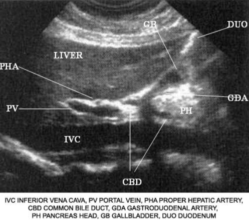
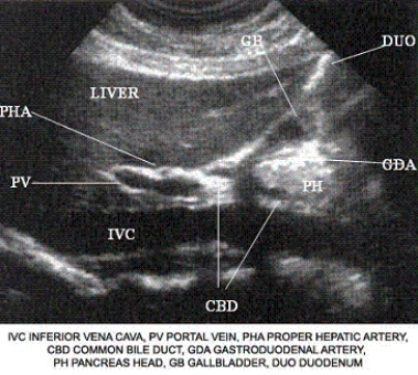
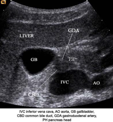
Describe the orientation of the major structures in this sagittal scanning plane image of the right upper quadrant.
 a. ________ section of the IVC
b. ________ section of the PV
c. ________ section of the CBD
d. ________ section of the PHA
e. ________ section of the PH
f. ________ section of the LIVER
a. ________ section of the IVC
b. ________ section of the PV
c. ________ section of the CBD
d. ________ section of the PHA
e. ________ section of the PH
f. ________ section of the LIVER
Free
(Short Answer)
4.9/5 (38)
Correct Answer:Verified
a. Longitudinal
b. Longitudinal
c. Longitudinal
d. Axial
e. Axial
f. Longitudinal
The IVC, this portion of the main portal vein, the common bile duct, and the liver are horizontally oriented. A sagittal image would demonstrate the length of these structures (longitudinal). The proper hepatic artery and head of the pancreas are vertically oriented. A sagittal image would demonstrate the axial plane of these structures.
Using the image below, describe the anatomic relationship of the common bile duct to adjacent structures.
(Essay)
4.9/5 (33)
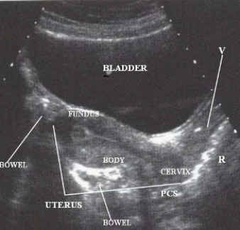
Using the image below, describe the anatomic relationship of the body of the uterus to adjacent structures.
(Essay)
4.8/5 (37)
Describe the orientation of the major structures in this sagittal scanning plane image just to the right of the midline of the body.
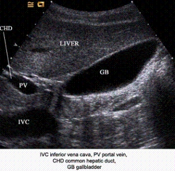 a. ____________ section of the IVC
b. ____________ section of the PV
c. ____________ section of the CHD
d. ____________ section of the GB
e. ____________ section of the liver
a. ____________ section of the IVC
b. ____________ section of the PV
c. ____________ section of the CHD
d. ____________ section of the GB
e. ____________ section of the liver
(Short Answer)
4.8/5 (38)
Which group of structures is located in the retroperitoneum?
(Multiple Choice)
4.8/5 (43)
Using the image below, describe the anatomic relationship of the inferior vena cava to adjacent structures.
(Essay)
4.9/5 (45)
Using the image below, describe the anatomic relationship of the hepatic artery to adjacent structures.
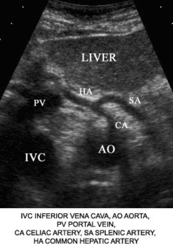
(Essay)
4.8/5 (27)
In gross anatomy, the tail of the pancreas is located ___________ to the splenic artery.
(Multiple Choice)
4.8/5 (36)
Describe the orientation of the major structures in this sagittal scanning plane image at the midline of the pelvis.
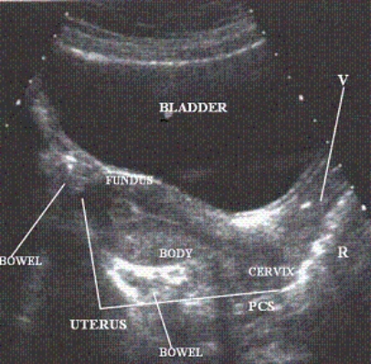 a. ________ section of the UTERUS
b. ________ section of the VAGINA
c. ________ section of the BLADDER
a. ________ section of the UTERUS
b. ________ section of the VAGINA
c. ________ section of the BLADDER
(Short Answer)
4.9/5 (39)
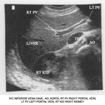
Describe the orientation of the major structures in this transverse scanning plane image of the right upper quadrant.
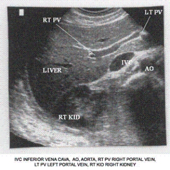 a. ________ section of the IVC
b. ________ section of the AO
c. ________ section of the RT KID
d. ________ section of the RT PV
e. ________ section of the LT PV
f. ________ section of the LIVER
a. ________ section of the IVC
b. ________ section of the AO
c. ________ section of the RT KID
d. ________ section of the RT PV
e. ________ section of the LT PV
f. ________ section of the LIVER
(Short Answer)
4.8/5 (44)
The single difference between structures seen on an ultrasound image section and a cadaver section is
(Multiple Choice)
4.7/5 (28)
Using the image below, describe the anatomic relationship of the portal vein to adjacent structures.
(Essay)
4.8/5 (34)
Using the image below, describe the anatomic relationship of the inferior vena cava to adjacent structures.
(Essay)
4.8/5 (35)
The renal arteries are located _________ to the renal veins.
(Multiple Choice)
4.8/5 (38)
Filters
- Essay(0)
- Multiple Choice(0)
- Short Answer(0)
- True False(0)
- Matching(0)