Exam 30: Three-Dimensional Ultrasound
Exam 1: Before and After the Ultrasound Examination30 Questions
Exam 2: Ultrasound Instrumentation: Knobology, Imaging Processing, and Storage25 Questions
Exam 3: General Patient Care10 Questions
Exam 4: First Scanning Experience35 Questions
Exam 5: Interdependent Body Systems40 Questions
Exam 6: Anatomy Layering and Sectional Anatomy40 Questions
Exam 7: Embryology50 Questions
Exam 8: The Abdominal Aorta30 Questions
Exam 9: The Inferior Vena Cava25 Questions
Exam 10: The Portal Venous System20 Questions
Exam 11: Abdominal Vasculature17 Questions
Exam 12: The Liver38 Questions
Exam 13: The Biliary System36 Questions
Exam 14: The Pancreas25 Questions
Exam 15: The Urinary System28 Questions
Exam 16: The Spleen20 Questions
Exam 17: The Gastrointestinal System35 Questions
Exam 18: The Prostate Gland and Seminal Vesicles17 Questions
Exam 19: The Female Pelvis31 Questions
Exam 20: First Trimester Obstetrics 0 to 12 Weeks29 Questions
Exam 21: Second and Third Trimester Obstetrics 13 to 42 Weeks25 Questions
Exam 22: High-Risk Obstetric Sonography23 Questions
Exam 23: The Thyroid and Parathyroid Glands32 Questions
Exam 24: Breast Sonography26 Questions
Exam 25: Penile and Scrotal Ultrasound20 Questions
Exam 26: The Neonatal Brain20 Questions
Exam 27: Pediatric Echocardiography20 Questions
Exam 28: Adult Echocardiography20 Questions
Exam 29: Vascular Technology30 Questions
Exam 30: Three-Dimensional Ultrasound32 Questions
Exam 31: Interventional and Intraoperative Ultrasound16 Questions
Select questions type
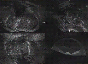
The main anatomy shown on this image is the __________.
Free
(Short Answer)
4.8/5  (30)
(30)
Correct Answer: Verified
prostate
The image demonstrates the prostate on 3D ultrasound.
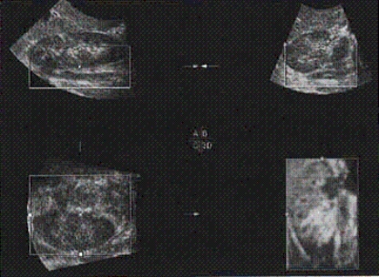
The main anatomy shown on this image is a(n) __________.
Free
(Short Answer)
4.9/5 (35)
Correct Answer:Verified
adult kidney
The image demonstrates an adult kidney on 3D ultrasound.
The SonoVCAD and SonoAVC are volumetric advances that can identify the
Free
(Multiple Choice)
4.8/5 (28)
Correct Answer:Verified
B
The acquisition of patient anatomy using 3D is called acquiring
(Multiple Choice)
5.0/5 (28)
The most important step in acquiring 3D ultrasound images is
(Multiple Choice)
4.8/5 (43)
Applications for three-dimensional (3D) ultrasound include which of the following?
(Multiple Choice)
4.8/5 (39)
What is the difference between Figure A and Figure B? (Hint: One is a recent scan; the other is much older. Can you tell which is which?)
(Essay)
4.9/5 (32)
Matrix array transducers represent advances in volume transducers. ____
(True/False)
4.9/5 (32)
Matrix array transducers contain hundreds of imaging elements. ____
(True/False)
4.7/5 (45)
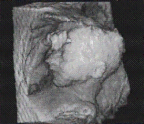
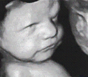
The rendering mode used for this fetal image is the __________?
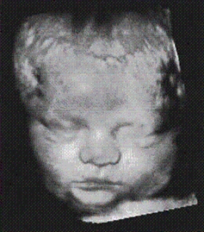
(Short Answer)
4.9/5 (37)
With automatic acquisition the "Region of Interest" box requires the sonographer to
(Multiple Choice)
4.9/5 (29)
Filters
- Essay(0)
- Multiple Choice(0)
- Short Answer(0)
- True False(0)
- Matching(0)