Exam 20: Exposure and Technique Errors
Exam 1: Radiation History10 Questions
Exam 2: Radiation Physics36 Questions
Exam 3: Radiation Characteristics24 Questions
Exam 4: Radiation Biology36 Questions
Exam 5: Radiation Protection28 Questions
Exam 6: Dental X-Ray Equipment13 Questions
Exam 7: Dental X-Ray Film46 Questions
Exam 8: Dental X-Ray Image Characteristics23 Questions
Exam 9: Dental X-Ray Film Processing51 Questions
Exam 10: Quality Assurance in the Dental Office27 Questions
Exam 11: Dental Radiographs and the Dental Radiographer13 Questions
Exam 12: Patient Relations and the Dental Radiographer10 Questions
Exam 13: Patient Education and the Dental Radiographer17 Questions
Exam 14: Legal Issues and the Dental Radiographer17 Questions
Exam 15: Infection Control and the Dental Radiographer30 Questions
Exam 16: Introduction to Radiographic Examinations16 Questions
Exam 17: Paralleling Technique35 Questions
Exam 18: Bisecting Technique36 Questions
Exam 19: Bite-Wing Technique20 Questions
Exam 20: Exposure and Technique Errors32 Questions
Exam 21: Occlusal and Localization Techniques20 Questions
Exam 22: Panoramic Imaging34 Questions
Exam 23: Extraoral Imaging27 Questions
Exam 24: Imaging of Patients With Special Needs19 Questions
Exam 25: Digital Imaging21 Questions
Exam 26: Three-Dimensional Digital Imaging24 Questions
Exam 27: Normal Anatomy: Intraoral Images75 Questions
Exam 28: Film Mounting and Viewing18 Questions
Exam 29: Normal Anatomy: Panoramic Images39 Questions
Exam 30: Introduction to Image Interpretation6 Questions
Exam 31: Descriptive Terminology18 Questions
Exam 32: Identification of Restorations, dental Materials, and Foreign Objects17 Questions
Exam 33: Interpretation of Dental Caries14 Questions
Exam 34: Interpretation of Periodontal Disease13 Questions
Exam 35: Interpretation of Trauma and Pulpal and Periapical Lesions20 Questions
Select questions type
When the third molar regions are not visible on a molar bite-wing image,the solution is to
Free
(Multiple Choice)
4.8/5  (38)
(38)
Correct Answer: Verified
D
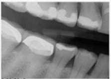
Identify the major technique error on your patient's bitewing.
Free
(Multiple Choice)
4.8/5 (38)
Correct Answer:Verified
D
To prevent underexposure,check and increase ___________ as needed.
1)exposure time
2)kilovoltage
3)milliamperage
Free
(Multiple Choice)
4.9/5 (34)
Correct Answer:Verified
A
Which of the following films would most likely appear black?
(Multiple Choice)
4.9/5 (41)
You have noticed that your mandibular incisors are foreshortened;how would you correct this problem?
(Multiple Choice)
4.8/5 (34)
Which of the following choices may be the cause of an unexposed film or receptor?
(Multiple Choice)
4.8/5 (41)
Your left premolar bite-wing image is lighter than the rest of the images and has a herringbone pattern.What can you do to prevent this problem from occurring? When the film is reversed,
(Multiple Choice)
4.9/5 (43)
The cause of an excessive margin of receptor edge (which appears as a black band)on a nondiagnostic periapical image is
(Multiple Choice)
4.9/5 (42)
The possibility of exposure of radiographic film to white light can be reduced by
(Multiple Choice)
4.8/5 (39)
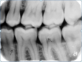
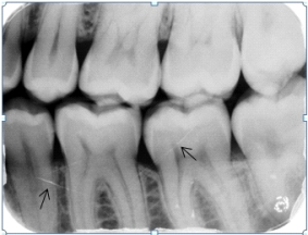
Identify the cause of the lines the arrows are pointing to on your patient's dental image.
(Multiple Choice)
4.8/5 (39)
The appearance of a patient's finger on the image is called a
(Multiple Choice)
4.7/5 (35)
The mesial half of your right premolar bite-wing image is clear,what will you need to correct when taking your retake to prevent this from reoccurring?
(Multiple Choice)
4.7/5 (30)
You have noticed that your maxillary premolars are elongated,how would you correct this problem?
(Multiple Choice)
4.8/5 (39)
Radiopaque artifacts or radiolucent scratch marks on the image indicate which of the following?
(Multiple Choice)
4.7/5 (34)
Filters
- Essay(0)
- Multiple Choice(0)
- Short Answer(0)
- True False(0)
- Matching(0)