Exam 34: Interpretation of Periodontal Disease
Exam 1: Radiation History10 Questions
Exam 2: Radiation Physics36 Questions
Exam 3: Radiation Characteristics24 Questions
Exam 4: Radiation Biology36 Questions
Exam 5: Radiation Protection28 Questions
Exam 6: Dental X-Ray Equipment13 Questions
Exam 7: Dental X-Ray Film46 Questions
Exam 8: Dental X-Ray Image Characteristics23 Questions
Exam 9: Dental X-Ray Film Processing51 Questions
Exam 10: Quality Assurance in the Dental Office27 Questions
Exam 11: Dental Radiographs and the Dental Radiographer13 Questions
Exam 12: Patient Relations and the Dental Radiographer10 Questions
Exam 13: Patient Education and the Dental Radiographer17 Questions
Exam 14: Legal Issues and the Dental Radiographer17 Questions
Exam 15: Infection Control and the Dental Radiographer30 Questions
Exam 16: Introduction to Radiographic Examinations16 Questions
Exam 17: Paralleling Technique35 Questions
Exam 18: Bisecting Technique36 Questions
Exam 19: Bite-Wing Technique20 Questions
Exam 20: Exposure and Technique Errors32 Questions
Exam 21: Occlusal and Localization Techniques20 Questions
Exam 22: Panoramic Imaging34 Questions
Exam 23: Extraoral Imaging27 Questions
Exam 24: Imaging of Patients With Special Needs19 Questions
Exam 25: Digital Imaging21 Questions
Exam 26: Three-Dimensional Digital Imaging24 Questions
Exam 27: Normal Anatomy: Intraoral Images75 Questions
Exam 28: Film Mounting and Viewing18 Questions
Exam 29: Normal Anatomy: Panoramic Images39 Questions
Exam 30: Introduction to Image Interpretation6 Questions
Exam 31: Descriptive Terminology18 Questions
Exam 32: Identification of Restorations, dental Materials, and Foreign Objects17 Questions
Exam 33: Interpretation of Dental Caries14 Questions
Exam 34: Interpretation of Periodontal Disease13 Questions
Exam 35: Interpretation of Trauma and Pulpal and Periapical Lesions20 Questions
Select questions type
The normal periodontal ligament space appears as a ______________ line.
Free
(Multiple Choice)
4.9/5  (38)
(38)
Correct Answer: Verified
B
Which of the following should you use to determine the extent of your patient's periodontal disease?
Free
(Multiple Choice)
4.9/5 (28)
Correct Answer:Verified
D
You suspect your patient has periodontal disease in the molar regions.Which of the following dental images should you take?
Free
(Multiple Choice)
4.7/5 (39)
Correct Answer:Verified
D
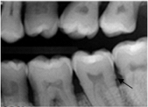
Identify the radiopaque area the arrow is pointing to on your patient's dental image.
(Multiple Choice)
4.9/5 (29)
In health,the lamina dura around the roots of the teeth appears as a(n)_________ line.
(Multiple Choice)
4.9/5 (30)
Dental images permit the evaluation of ______ in the detection of periodontal disease.
(Multiple Choice)
4.9/5 (35)
The periodontal ligament space is located between the root of the tooth and the
(Multiple Choice)
4.8/5 (38)
Your patient is viewing her bite-wing images and has noticed the small radiopaque pieces on the interproximal surfaces below the contacts of some of her teeth.What is she looking at?
(Multiple Choice)
4.8/5 (39)
A patient with localized bone loss will exhibit bone loss in less than ______ of the areas.
(Multiple Choice)
4.9/5 (39)
Your patient has been diagnosed as having a periodontal classification of ADA case type I.How far below the cement-enamel junction would you expect to see the crestal bone on the dental images?
(Multiple Choice)
4.9/5 (45)
The _________________ of adjacent teeth can be used as a plane of reference in determining the pattern of bone loss present.
(Multiple Choice)
4.8/5 (39)
Filters
- Essay(0)
- Multiple Choice(0)
- Short Answer(0)
- True False(0)
- Matching(0)