Exam 1: Cellular Responses to Injury
Exam 1: Cellular Responses to Injury5 Questions
Exam 2: Acute Inflammation, Healing and Repair13 Questions
Exam 3: Chronic Inflammation9 Questions
Exam 4: Infections of Histological Importance18 Questions
Exam 5: Amyloidosis3 Questions
Exam 6: Disorders of Growth3 Questions
Exam 7: Dysplasia and Neoplasia2 Questions
Exam 8: Atherosclerosis8 Questions
Exam 9: Thrombosis and Embolism7 Questions
Exam 10: Infarction9 Questions
Exam 11: Cardiovascular System12 Questions
Exam 12: Respiratory System11 Questions
Exam 13: Gastrointestinal System8 Questions
Exam 14: Liver and Pancreaticobiliary System8 Questions
Exam 15: Urinary System9 Questions
Exam 16: Lymphoid and Haematopoietic Systems6 Questions
Exam 17: Female Reproductive System7 Questions
Exam 18: Breast7 Questions
Exam 19: Male Reproductive System2 Questions
Exam 20: Endocrine System4 Questions
Exam 21: Skin8 Questions
Exam 22: Bone and Soft Tissues6 Questions
Exam 23: Nervous System8 Questions
Select questions type
The structure designated by the "V" in the accompanying photomicrograph of liver is formed by which one of the following pathophysiologic mechanisms? 
Free
(Multiple Choice)
4.9/5  (32)
(32)
Correct Answer: Verified
D
A renal glomerulus (G)and surrounding tubules are shown in the accompanying photomicrograph.Which one of the following is the best description of the histopathology seen? 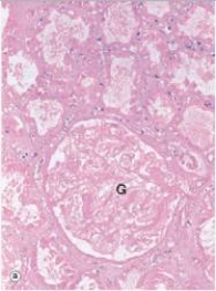
Free
(Multiple Choice)
4.8/5 (32)
Correct Answer:Verified
A
The structure labelled "A" in the accompanying photomicrograph of colonic mucosa represents which one of the following processes? 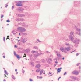
Free
(Multiple Choice)
4.8/5 (34)
Correct Answer:Verified
A
The structure designated by the "V" in the accompanying photomicrograph of liver likely contains which one of the following substances during life? 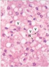
(Multiple Choice)
4.9/5 (38)
The nuclei in the liver cells designated with an "N" in the accompanying photomicrograph appear abnormal.Which one of the following terms best describes their appearance? 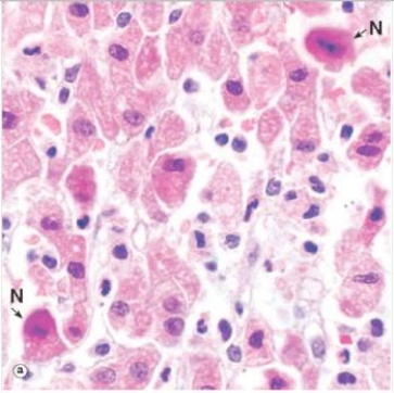
(Multiple Choice)
4.8/5 (35)
Filters
- Essay(0)
- Multiple Choice(0)
- Short Answer(0)
- True False(0)
- Matching(0)