Exam 23: Nervous System
Exam 1: Cellular Responses to Injury5 Questions
Exam 2: Acute Inflammation, Healing and Repair13 Questions
Exam 3: Chronic Inflammation9 Questions
Exam 4: Infections of Histological Importance18 Questions
Exam 5: Amyloidosis3 Questions
Exam 6: Disorders of Growth3 Questions
Exam 7: Dysplasia and Neoplasia2 Questions
Exam 8: Atherosclerosis8 Questions
Exam 9: Thrombosis and Embolism7 Questions
Exam 10: Infarction9 Questions
Exam 11: Cardiovascular System12 Questions
Exam 12: Respiratory System11 Questions
Exam 13: Gastrointestinal System8 Questions
Exam 14: Liver and Pancreaticobiliary System8 Questions
Exam 15: Urinary System9 Questions
Exam 16: Lymphoid and Haematopoietic Systems6 Questions
Exam 17: Female Reproductive System7 Questions
Exam 18: Breast7 Questions
Exam 19: Male Reproductive System2 Questions
Exam 20: Endocrine System4 Questions
Exam 21: Skin8 Questions
Exam 22: Bone and Soft Tissues6 Questions
Exam 23: Nervous System8 Questions
Select questions type
A 67 year old man,who had been an accounting executive for a large manufacturing firm most of his career,began to have problems driving in his city.He frequently became lost.He was also having difficulties finding the proper word to describe simple things,like a fork,or a pair of pliers.A CT scan showed atrophy of cerebral cortex,and a relative increase in size of the lateral ventricles.He died of an apparent heart attack a few months after the onset of symptoms,and his wife requested an autopsy.Representative sections are shown in the accompanying photomicrographs.Which one of the following is the best diagnosis? 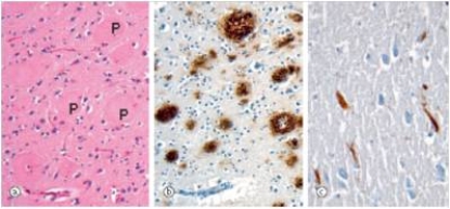
Free
(Multiple Choice)
4.8/5  (37)
(37)
Correct Answer: Verified
A
The wife of a 55 year old man noticed that he exhibited strange behaviour such as fits of rage and moments of confusion.Together,they consulted a GP who also noticed some neurologic abnormalities such as rigidity in the upper extremities.A representative section of the brain biopsy is shown in the accompanying photomicrograph.Which one of the following is the best diagnosis? 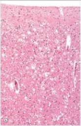
Free
(Multiple Choice)
4.8/5 (34)
Correct Answer:Verified
E
A 44 year old man began exhibiting bizarre personality changes,noted first by his wife.She brought him to the local Emergency Department,where a neuropsychiatrist ordered a CT scan.The scan revealed a 3 cm,well-demarcated tumour in the frontal lobes.Calcification was evident within the tumour.A neurosurgeon was consulted,and the tumour was later removed without incident.The patient returned to his normal state.A representative section of the tumour is shown in the accompanying photomicrograph.Which one of the following is the best diagnosis? 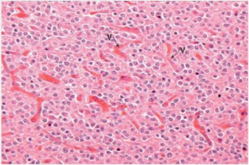
Free
(Multiple Choice)
4.9/5 (34)
Correct Answer:Verified
E
A 47 year old woman consulted her physician because of frequent headaches and weakness in her left arm.A CT scan showed a large 4 cm tumour on the right convexity,attached to the dura.It compressed,but did not invade the motor cortex.The tumour was easily removed.A representative section from the tumour is shown in the accompanying photomicrograph.Which one of the following is the best diagnosis? 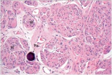
(Multiple Choice)
4.9/5 (39)
A 37 year old man had suffered from hemiplegia for several years.He had been told it was multiple sclerosis.A neurologist suspected that the diagnosis was incorrect,and ordered a CT scan of the patient's cervical spine.The scan revealed a central tumour,which was pressing on the corticospinal tract.A neurosurgeon was able to completely remove the tumour,which is shown in the accompanying photomicrograph.Which one of the following is the best diagnosis? 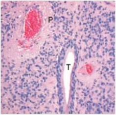
(Multiple Choice)
4.9/5 (32)
A 45-year-old woman woke up in the morning with a severe headache.She did not recognize her husband of 20 years.She then experienced a grand mal seizure.Her husband called an ambulance,and she was taken to the hospital.A CT scan disclosed a large tumour,deep in the cerebrum,that crossed the midline near the corpus callosum.Necrosis was evident throughout the tumour.A brain biopsy of a portion of the tumour was done and a representative section is shown in the accompanying photomicrograph.Which one of the following is the best diagnosis? 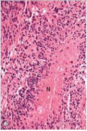
(Multiple Choice)
4.9/5 (39)
A 3 year old child was having difficulty walking.He had not met his developmental milestones.His legs actually looked quite strong,with apparently muscular calves.However,the paediatric neurologist ordered a muscle biopsy,which is shown in the accompanying photomicrograph.On the right is an immunohistochemical stain for dystrophin protein. 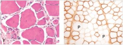
(Multiple Choice)
4.9/5 (38)
A 32 year old woman suddenly developed paraplegia in her right leg.This caused her to crash her car.A coroner's autopsy was ordered.A representative section from the area of the internal capsule in the brain disclosed the findings seen in the accompanying photomicrographs,with H&E in the upper photo (a)and a Luxol fast blue myelin stain in the lower photo (c).Which one of the following is the best diagnosis? 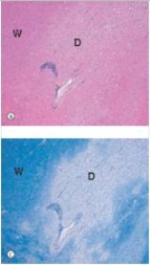
(Multiple Choice)
4.9/5 (31)
Filters
- Essay(0)
- Multiple Choice(0)
- Short Answer(0)
- True False(0)
- Matching(0)